

doi: 10.1126/science.1112766.
Epub 2005 Aug 25.
Suppression of aging in mice by the hormone Klotho
Affiliations
- PMID: 16123266
- PMCID: PMC2536606
- DOI: 10.1126/science.1112766
Item in Clipboard
Suppression of aging in mice by the hormone Klotho
Science.
.
Abstract
A defect in Klotho gene expression in mice accelerates the degeneration of multiple age-sensitive traits. Here, we show that overexpression of Klotho in mice extends life span. Klotho protein functions as a circulating hormone that binds to a cell-surface receptor and represses intracellular signals of insulin and insulin-like growth factor 1 (IGF1), an evolutionarily conserved mechanism for extending life span. Alleviation of aging-like phenotypes in Klotho-deficient mice was observed by perturbing insulin and IGF1 signaling, suggesting that Klotho-mediated inhibition of insulin and IGF1 signaling contributes to its anti-aging properties. Klotho protein may function as an anti-aging hormone in mammals.
Figures
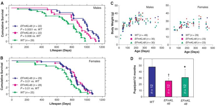
Klotho overexpression extends life span in the mouse. Kaplan-Meier analysis of survival in (A) males [P = 0.006 in EFmKL46 versus wild-type (WT) mice, and P < 0.0001 in EFmKL48 versus wild type by log-rank test) and in (B) females (P = 0.01 in EFmKL46 versus wild type, and P = 0.01 in EFmKL48 versus wild type by log-rank test). The average life span of male wild-type, EFmKL46, and EFmKL48 mice was 715 ± 44 days, 858 ± 40 days, and 936 ± 47 days (means ± SEM), respectively. The average life span of female wild-type, EFmKL46, and EFmKL48 mice was 697 ± 45 days, 829 ± 32 days, and 830 ± 29 days, respectively. (C) Body weight of wild-type, EFmKL46, and EFmKL48 mice. No significant difference in growth was observed. (D) Klotho overexpression reduces fecundity. Twelve breeding pairs at 12 weeks of age were set up for each genotype. The number of offspring generated during 12 months was recorded for each breeding pair. Although average litter size (pups per birth) of wild-type, EFmKL46, and EFmKL48 pairs was not significantly different (6.6 ± 1.0, 6.1 ± 1.3, and 7.0 ± 1.2, respectively), the number of births (births per pair per 12 months) was fewer in transgenic mice pairs (7.2 ± 1.6, 4.2 ± 0.8, and 4.5 ± 2.2, respectively), resulting in significantly fewer offspring in transgenic pairs. Data are means ± SD. *, P < 0.05; †, P < 0.01 versus wild-type mice by analysis of variance (ANOVA).
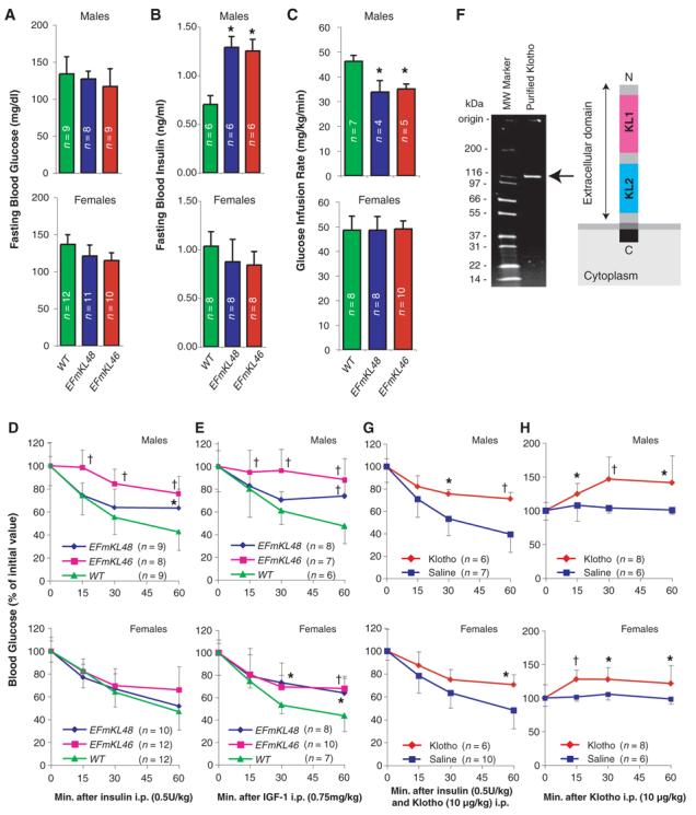
Klotho increases resistance to insulin and IGF1. (A and B) Fasting blood glucose (A) and insulin (B) levels were compared between wild-type (WT), EFmKL48, and EFmKL46 mice. (C) Hyperinsulinemic euglycemic clamp experiments. Glucose infusion rate (mg/kg/min) was compared between wild-type, EFmKL48, and EFmKL46 mice. During the clamp experiments there were no differences in blood glucose concentration between wild-type, EFmKL46, and EFmKL48 mice. The number of animals (n) for each group is indicated in the bars. (D and E) Insulin (D) and IGF1 (E) tolerance tests. Blood glucose levels after injection of insulin (0.5 U/kg) or IGF1 (0.75 mg/kg) were expressed as a percentage change from blood glucose concentration at time zero. Error bars indicate SD. *, P < 0.05 and †, P < 0.01 versus wild-type mice by ANOVA. (F) A schematic representation of Klotho extracellular peptide (right) and analysis of purified recombinant Klotho peptide by SDS-polyacrylamide gel electrophoresis (left). (G) The effect of Klotho injection on insulin tolerance in mice. Insulin tolerance tests were performed with age-matched wild-type mice immediately after intraperitoneal injection with saline or purified recombinant Klotho peptide (10 μg/kg). (H) The effect of Klotho injection on blood glucose levels. Saline or Klotho protein (10 μg/kg) was administered into age-matched wild-type mice by intraperitoneal injection. Error bars indicate SD. *, P < 0.05 and †, P < 0.01 versus saline-injected mice by ANOVA.
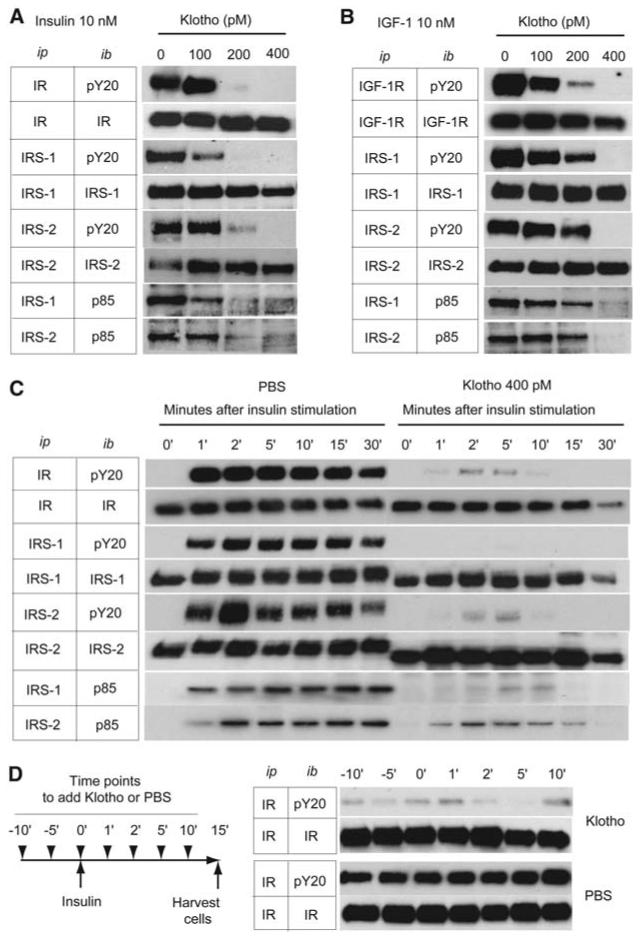
Klotho protein inhibits intracellular insulin and IGF1 signaling. (A and B) Effect of Klotho on tyrosine phosphorylation of insulin and IGF1 receptors as well as IRS-1 and IRS-2, association of IRS-1 and IRS-2 with the PI3-kinase regulatory subunit (p85), and phosphorylation of Akt in L6 cells stimulated with 10 nM of insulin (A) or 10 nM of IGF1 (B). Antibodies used for immunoprecipitation (ip) and immunoblotting (ib) were indicated. IR, antibody to insulin receptor β chain; pY20, antibody to phosphotryrosine; IRS-1, antibody to IRS-1; IRS-2, antibody to IRS-2; p85, antibody to PI3-kinase regulatory subunit; IGF-1R, antibody to IGF1 receptor β chain. (C) A time course of the inhibitory effect of Klotho protein (400 pM) on insulin signaling in H4IIE cells. The cells were harvested before (0′) and at the indicated time points after insulin stimulation (10 nM). (D) Inactivation of activated insulin receptor by Klotho protein. H4IIE cells were stimulated with insulin (10 nM) at time 0′ and harvested 15 min later. Klotho (400 pM) or phosphate-buffered saline (PBS) was added at the indicated time points indicated (left panel). The cell lysates were immunoprecipitated with IR and immunoblotted with pY20 or IR (right panel).
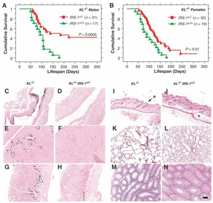
Rescue of aging-like phenotypes in KL−/− mice by genetic intervention in insulin and IGF1 signaling. (A and B) Life-span extension in KL−/− mice by reducing IRS-1 expression. KL−/− mice heterozygous for an IRS-1 null allele (IRS-1+/−) lived longer than those without the mutation (IRS-1+/+) both in males (P = 0.0005 by log-rank test) and females (P = 0.01 by log-rank test). [(C) to (N)] Rescue of aging-like phenotypes in KL−/− IRS-1+/− mice at the histological level. Typical findings from four 8-week-old males of each genotype are shown. (C and D) Aorta (von Kossa staining). Calcification of arterial walls [black deposits in (C)] was decreased in KL−/− IRS-1+/− mice (D). (E and F) Kidney (von Kossa staining). Calcification of small arteries and renal tubules [black deposits in (E)] was decreased in KL−/− IRS-1+/− mice (F). (G and H) Stomach (von Kossa staining). Ectopic calcification in gastric mucosa and small arteries [black deposits in (G)] was alleviated in KL−/− IRS-1+/− mice (H). (I and J) Cross-sections of the skin. Hematoxylin-eosin (HE) staining. Reduction in epidermal layer (e) thickness observed in KL−/− mice (I) was improved and subcutaneous fat (s) was restored in KL−/− IRS-1+/− mice (J). (K and L) Lung (HE staining). Emphysematous changes, including enlargement of air spaces and destruction of the normal alveolar architecture were observed in KL−/− mice (K), but were alleviated in KL−/− IRS-1+/− mice (L). (M and N) Testis (HE staining). Seminiferous tubules were atrophic and no mature sperm was observed in KL−/− mice (M). Spermatogenesis was restored in KL−/− IRS-1+/− mice (N). All panels were shown in the identical magnification (×200). Scale bar, 200 μm.
References
Publication types
MeSH terms
Substances
Grants and funding
LinkOut - more resources
Full Text Sources
Other Literature Sources
Medical
Molecular Biology Databases
Miscellaneous
