. 2006 Feb;24(2):274-83.
doi: 10.1634/stemcells.2005-0059. Epub 2005 Aug 25.

Embryonic stem cell-derived neural progenitors incorporate into degenerating retina and enhance survival of host photoreceptors

Affiliations

Embryonic stem cell-derived neural progenitors incorporate into degenerating retina and enhance survival of host photoreceptors

Jason S Meyer et al. Stem Cells. 2006 Feb.

Abstract

Embryonic stem (ES) cells differentiate into all cell types of the body during development, including those of the central nervous system (CNS). After transplantation, stem cells have the potential to replace host cells lost due to injury or disease or to supply host tissues with therapeutic factors and thus provide a functional benefit. In the current study, we assessed whether mouse neuralized ES cells can incorporate into retinal tissue and prevent retinal degeneration in mnd mice. These mice have an inherited lysosomal storage disease characterized by retinal and CNS degeneration. Sixteen weeks after intravitreal transplantation into adult mice, donor cells had incorporated into most layers of the retina, where they resembled retinal neurons in terms of morphology, location in the retina, and expression of cell type-specific marker proteins. Presence of these donor cells was correlated with a reduction in the sizes and numbers of lysosomal storage bodies in host retinal cells. The presence of transplanted donor cells was also accompanied by enhanced survival of host retinal neurons, particularly photoreceptors. These results demonstrate that neuralized ES cells protect host neurons from degeneration and appear to replace at least some types of lost neurons.


Conflict of interest statement

Disclosures

The authors indicate no potential conflicts of interest.

Figures

Figure 1
Figure 1
Donor cells become associated with the host retina after intravitreal transplantation. (A–H): Images of the whole-mount retina preparation displayed in the whole-mount montage shown in the center. The locations from which these images were taken are indicated in the montage. Many enhanced green fluorescent protein-expressing donor cells become widely dispersed across the retina and adopt neural-like morphologies. Scale bar = 200 μm.
Figure 2
Figure 2
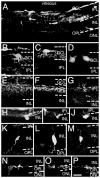
Donor cells incorporate within many retinal layers of mnd mice and acquire retinal cell–like morphologies. Transplanted cells expressing enhanced green fluorescent protein are found integrated within the inner layers and outer plexiform layer of the host retina and are widely distributed by 16 weeks after transplantation (A). Retinal cell-like morphologies are seen in most layers, including donor cells that resemble ganglion cells (B–D), extensive branching within the inner plexiform layers (E–G), amacrine cell–like cells (H–J), bipolar cell–like cells (K–M), and cells that appear to be horizontal cells (N–P). Scale bar = 80 μm (A) and 40 μm (B–P). Abbreviations: GCL, ganglion cell layer; INL, inner nuclear layer; IPL, inner plexiform layer; ONL, outer nuclear layer; OPL, outer plexiform layer.
Figure 3
Figure 3
Transplanted cells express neural- and retinal-specific markers in vivo. Donor cells found throughout the retina display general and specific neural markers. (A–F): Transplanted cells acquire neuronal-like morphologies and express general neural markers, such as β-III tubulin (A–C) and NeuN (D–F). (G–L): Transplanted cells with retinal cell–like morphologies can express specific markers appropriate for cells unique to particular layers, such as the amacrine cell marker calretinin (G–I) and the bipolar cell marker cPKC-α (J–L). Donor cells immediately adjacent to the outer nuclear layer often express the photoreceptor marker rhodopsin (M–O). Note that cells expressing specific markers possess morphologies appropriate for the corresponding retinal cell types found in these layers of the host retina, such as the bipolar-like cell in (J). (P–R): Neurite-like processes expressing enhanced green fluorescent protein are found largely within the plexiform layers and express the synaptic vesicle marker SV2. (S–U): The astrocyte marker GFAP was expressed by some donor cells. The GFAP expression was typically found in donor cells remaining on the vitreal surface of the retina. Note that some host reactive Müller cells expressed GFAP. The panels shown here represent merged images, i.e., images captured using filters specific for either GFP or rhodamine fluorescence. The respective images were given false colors (GFP, green and rhodamine; red) and then merged using Photoshop. Therefore, cell bodies and processes that appear yellow in the merged images exhibited fluorescence due to both enhanced GFP expression and labeling for the indicated marker. Scale bar = 40 μm. Abbreviation: GFP, green fluorescent protein.
Figure 4
Figure 4
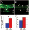
The presence of donor cells reduces lysosomal storage bodies in host retina. Autofluorescent lysosomal storage bodies (arrowheads) are a signature feature of the neuronal ceroid lipofuscinoses. The yellow storage body autofluorescence and enhanced green fluorescent protein fluorescence are simultaneously visible when viewed with fluorescein isothiocyanate filters. (A, B): Lysosomal storage bodies are less abundant in mnd retinal samples in which donor cells are present at 16 weeks after transplantation (A) compared with age-matched sham-injected controls (B). (C): The mean number of storage bodies is significantly less in samples with donor cell incorporation (Cells Present) compared with sham-injected controls (Cells Absent). (D): Likewise, the mean two-dimensional surface area of lysosomal storage bodies in host retinas is significantly smaller in samples exhibiting donor cell incorporation compared with sham-injected controls. *p < .01 (C, D). Scale bar = 50 μm (A, B). Abbreviations: GCL, ganglion cell layer; INL, inner nuclear layer; IPL, inner plexiform layer; ONL, outer nuclear layer; OPL, outer plexiform layer.
Figure 5
Figure 5
Donor cells rescue retinal photoreceptors from cell death. The primary cell type lost due to the degenerative process in the mnd retina is the photoreceptor. (A): In sham-injected, age-matched controls, the few photoreceptors (rhodopsin labeling is blue) remaining have fragmented outer segments and typically the retina contains only one or two layers of photoreceptor cell bodies, as revealed by nuclear labeling with propidium iodide (red). (B): At 16 weeks after transplantation, substantial numbers of photoreceptors with intact outer segments remain in host retinas that contain donor cells (green). (C): A normal, age-matched C57 retina. (D): Significantly more host photoreceptors (cells in ONL expressing Rhodopsin [i.e., Rhodopsin +]; nuclei of these cells were counted) survive in the presence of donor cells (Cells Present) in both central retina (Central) and peripheral retina (Peripheral) (*p < .01). The scale bar = 50 μm (C) and 25 μm (A, B). Abbreviations: GCL, ganglion cell layer; INL, inner nuclear layer; IPL, inner plexiform layer; ONL, outer nuclear layer; OPL, outer plexiform layer.
Figure 6
Figure 6
Donor cells exert regional rescue effects upon host tissue. Reduction of storage body size and number, as well as rescue of host photoreceptors, was greater in close association with integrated donor cells. (A): Storage body number was significantly decreased in close association with donor cells within the same retinal samples. (B): Likewise, the mean storage body size was significantly reduced in close association with donor cells within the same retinal samples. (C): Rescue of host photoreceptors was significantly enhanced in close association with integrated donor cells. *p < .05 (A–C).

References

    1. Brook FA, Gardner RL. The origin and efficient derivation of embryonic stem cells in the mouse. Proc Natl Acad Sci U S A. 1997;94:5709–5712. - PMC - PubMed
    1. Robertson EJ. Derivation and maintenance of embryonic stem cell cultures. Methods Mol Biol. 1997;75:173–184. - PubMed
    1. Smith AG. Embryo-derived stem cells: Of mice and men. Annu Rev Cell Dev Biol. 2001;17:435– 462. - PubMed
    1. Itskovitz-Eldor J, Schuldiner M, Karsenti D, et al. Differentiation of human embryonic stem cells into embryoid bodies compromising the three embryonic germ layers. Mol Med. 2000;6:88–95. - PMC - PubMed
    1. Lumelsky N, Blondel O, Laeng P, et al. Differentiation of embryonic stem cells to insulin-secreting structures similar to pancreatic islets. Science. 2001;292:1389–1394. - PubMed

Publication types