. 2005 Sep;167(3):637-49.
doi: 10.1016/S0002-9440(10)62039-1.

Blocking of monocyte chemoattractant protein-1 during tubulointerstitial nephritis resulted in delayed neutrophil clearance

Affiliations

Blocking of monocyte chemoattractant protein-1 during tubulointerstitial nephritis resulted in delayed neutrophil clearance

Ping Li et al. Am J Pathol. 2005 Sep.

Abstract

The chemokine monocyte chemoattractant protein (MCP)-1 has been implicated in the monocyte/macrophage infiltration that occurs during tubulointerstitial nephritis (TIN). We investigated the role of MCP-1 in rats with TIN by administering a neutralizing anti-MCP-1 antibody (Ab). We observed significantly reduced macrophage infiltration and delayed neutrophil clearance in the kidneys of TIN model rats treated with the anti-MCP-1 Ab. To exclude the possibility that an observed immune complex could affect the resolution of apoptotic neutrophils via the Fc receptor, TIN model rats were treated with a peptide-based MCP-1 receptor antagonist (RA). The MCP-1 RA had effects similar to those of the anti-MCP-1 Ab. In addition, MCP-1 did not affect macrophage-mediated phagocytosis of neutrophils in vitro. Deposition of the anti-MCP-1 Ab in rat kidneys resulted from its binding to heparan sulfate-immobilized MCP-1, as demonstrated by the detection of MCP-1 in both pull-down and immunoprecipitation assays. We conclude that induction of chemokines, specifically MCP-1, in TIN corresponds with leukocyte infiltration and that the anti-MCP-1 Ab formed an immune complex with heparan sulfate-immobilized MCP-1 in the kidney. Antagonism of MCP-1 in TIN by Ab or RA may alter the pathological process, most likely through delayed removal of apoptotic neutrophils in the inflammatory loci.


Figures

Figure 1
Figure 1
Characterization of Abs against MIP-2, KC, and MCP-1. Expression of MIP-2 (a), KC (b), and MCP-1 (c) was detected in transfectants using rabbit anti-rat polyclonal Abs generated from recombinant proteins expressed in E. coli. Plasmids (20 μg), expressing full-length chemokine cDNA, were transfected into the human kidney embryo cell line 293T using electroporation. After 72 hours of culture, supernatants were collected and analyzed by Western blot: lane 1, MIP-2 transfectant; lane 2, KC transfectant; lane 3, MCP-1 transfectant. Expression of MIP-2 (left), KC (middle), and MCP-1 (right) was observed in the transfectants. d: Coomassie blue staining of purified MCP-1 (full length), MCP-1 RA(9-73), and MCP-1(11-73). Recombinant MCP-1 analogues were generated in E. coli. Purification was performed by affinity chromatography using a nickel column. Left lane: protein molecular weight markers; lane 1, 1 μg of MCP-1(11-73); lane 2, 1 μg of MCP-1RA(9-73); lane 3: 1 μg of MCP-1 (full length).
Figure 2
Figure 2
Evaluation of peritoneal macrophage migration mediated by MCP-1 and effect of anti-MCP-1 Ab and MCP-1 RA on MCP-1-mediated chemotaxis in vitro. Recombinant rat MCP-1 mediated the migration of peritoneal macrophages in a dose-dependent manner. Anti-MCP-1(1-73) Ab (a) and MCP-1 RA(9-73) (b) inhibit chemotaxis of peritoneal macrophage induced by MCP-1. Control experiments were performed with preimmune rabbit serum and MCP-1(11-73) peptide. Each graph shown represents one set of experiments (n = 3). All results were verified on two further occasions. The total number of migrating cells in five high-power fields is indicated on the y axis (mean ± SD).
Figure 3
Figure 3
Assessment of MCP-1, MIP-2, and KC expression in the TIN model kidney by RPA and Western blotting. Experiments were conducted following the protocol mentioned in the Materials and Methods section. a and b: Renal total RNA (6 μg) was used for each assay. Left lane of each panel contains probes for MCP-1 (239 bp), MIP-2 (174 bp), and KC (274 bp). Housekeeping gene L32 was used as a loading control. The time points (days after injection of bovine tubular basement membrane) are marked. The protected bands are shorter than the probes due to the unhybridized sequences in the polylinker regions of the probes were digested by RNase A and T1, and undigested KC probe is marked with an asterisk. The data shown are representative of three independent experiments. c: Immunoblots assessing chemokine expression (MCP-1, MIP-2, and KC) in homogenates from the TIN model kidney.
Figure 4
Figure 4
The effects of anti-MCP-1(1-73) Ab (c, d) and MCP-1 RA(9-73) peptide (g, h) on neutrophil and macrophage infiltration in rat TIN model kidney (10 days). Neutrophils were identified (left, red staining) using the anti-MPO Ab. Macrophages were identified (right, brown staining) using the ED1 Ab. Sham animals treated with NRS exhibit a high degree of macrophage infiltration (b) and substantial neutrophil clearance (a) in the area of inflammation; whereas, animals treated with anti MCP-1(1-73) Ab exhibit decreased macrophage infiltration (d) and a concomitant accumulation of vast numbers of neutrophils (c) in the injured tissue. Animals treated with the MCP-1 RA(9-73) peptide exhibit a similar delay in neutrophil clearance (g) corresponding to a low degree of macrophage infiltration (h) in the area of injury; whereas, animals treated with an inactive form of the MCP-1(11-73) peptide exhibit neutrophil clearance (e) and macrophage infiltration (f) similar to sham animals. The number of MPO+ and ED1+ cells were calculated in 10 fields of tubulointerstitial area in each sample (i, j). Original magnifications, ×200.
Figure 5
Figure 5
Immunofluorescent staining of C3 in kidneys of TIN rats treated with anti-MCP-1(1-73) Ab or NRS. The level of complement component C3 deposition in the anti-MCP-1(1-73) Ab-treated TIN model kidney. Rat kidneys were processed with cryosectioning (0.5 μm), followed by probing with fluorescein isothiocyanate-conjugated rabbit anti-rat C3 Ab, which recognizes an epitope expressed by C3. Immunofluorescent staining revealed that C3 protein was localized on the outer surface of the proximal and distal tubules in both NRS-treated group (a) and in anti-MCP-1 Ab-treated animals (b). Original magnifications, ×200.
Figure 6
Figure 6
The process of phagocytosis of macrophages in TIN model rats treated with anti-MCP-1(1-73) Ab. A and B: Phagocytosis in vivo. Consecutive sections of rat kidneys with TIN were stained either for neutrophils, using the anti-MPO Ab (left), or for monocytes/macrophages using the ED1 Ab (right). Neutrophil staining is intensely purple while macrophage staining is dark brown. Top: A and B demonstrate the fully formed (area marked with asterisk) or partially formed (areas labeled with a, a’, b, and b’) multinuclear giant cells as macrophages engulf and ingest neutrophils in the area of inflammatory loci. Bottom: a, a’, b, and b’ illustrate this process at higher magnification, with neutrophils that are being engulfed indicated by arrows. c and d: Phagocytosis in vitro. Neutrophils within macrophages are readily identifiable by positive staining (brown) for MPO. Two representative images of macrophages treated with vehicle (a) or MCP-1 (b). The experiment was done three times. Original magnifications: ×100 (A, B); ×200 (ad).
Figure 7
Figure 7
RNase protection analysis of cytokine mRNA expression in the kidney of rats treated with anti-MCP-1 Ab or NRS. Each lane represents a single rat sampled. Probes contain polylinker regions and are longer than the protected bands. Rat ribosomal L32 and GAPDH genes were used as a housekeeping gene.
Figure 8
Figure 8
Representative Masson’s trichrome staining of renal sections from a normal rat (a), TIN rat (b), anti-MCP-1 Ab-treated rat (c), and MCP-1 RA-treated rat (d). Three dyes are used selectively to stain collagen (blue), nuclei (black), and background (red). Original magnifications, ×200.
Figure 9
Figure 9
Comparison of rabbit IgG and HS distribution in TIN model rats. a and b: Localization of rabbit IgG in the anti-MCP-1(1-73) Ab-treated TIN model kidney. Rabbit IgG was directly detected by a horseradish peroxidase-labeled goat anti-rabbit Ab in NRS-treated group (a) and anti MCP-1(1-73) Ab-treated group (b). c and d: Induction of HS in the TIN model. a: Rat kidney stained with anti-HS Ab against 10E4 epitope shows that the expression of HS is mainly located to the lumen of the proximal and distal tubules and to Bowman’s capsule of the glomeruli. b: TIN model rat kidney shows extensive staining of HS in the vascular endothelial cells as well as in the Bowman’s capsule of the glomeruli and in the epithelial cells of the interstitial tubules. Numerous infiltrated inflammatory cells in the interstitial lesions were observed when counterstaining (blue) was performed. Original magnifications, ×200.
Figure 10
Figure 10
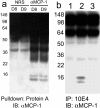
Protein interaction between MCP-1, rabbit anti-MCP-1 Ab, and HS. The immune complex of MCP-1 and rabbit anti-MCP-1 Ab was detected using pull-down and immunoprecipitation assays (left). Left: Pull-down experiments were performed using protein A followed by immunoblotting for MCP-1. Kidney homogenates from TIN models day 8 and day 9 were subjected to pull-down using protein A beads and were tested for the presence of MCP-1 by rabbit anti-rat MCP-1 Ab. Right: Immunoprecipitation was performed using mouse anti-rat HS Ab 10E4 followed by immunoblotting for MCP-1. Kidney cell lysate from normal and TIN model kidneys were subjected to immunoprecipitation using the 10E4 Ab (lanes 1 and 2). A sample of cell lysate from TIN model kidney was subjected to digestion with heparanase (lane 3) after the immunoprecipitation with 10E4. An immunoblot for MCP-1 was then performed on all three samples.

Similar articles

Cited by

References

    1. Zanetti M, Wilson CB. Characterization of anti-tubular basement membrane antibodies in rats. J Immunol. 1983;130:2173–2179. - PubMed
    1. Mampaso FM, Wilson CB. Characterization of inflammatory cells in autoimmune tubulointerstitial nephritis in rats. Kidney Int. 1983;23:448–457. - PubMed
    1. Ryan GB, Majno G. Acute inflammation. A review. Am J Pathol. 1977;86:183–276. - PMC - PubMed
    1. Baggiolini M, Dewald B, Moser B. Human chemokines: an update. Annu Rev Immunol. 1997;15:675–705. - PubMed
    1. Baggiolini M, Moser B, Clark-Lewis I. Interleukin-8 and related chemotactic cytokines. The Giles Filley Lecture. Chest. 1994;105:95S–98S. - PubMed

Publication types

MeSH terms

LinkOut - more resources