

Transcriptional up-regulation and activation of initiating caspases in experimental glaucoma
- PMID: 16127148
- PMCID: PMC1698740
- DOI: 10.1016/S0002-9440(10)62042-1
Transcriptional up-regulation and activation of initiating caspases in experimental glaucoma
Abstract
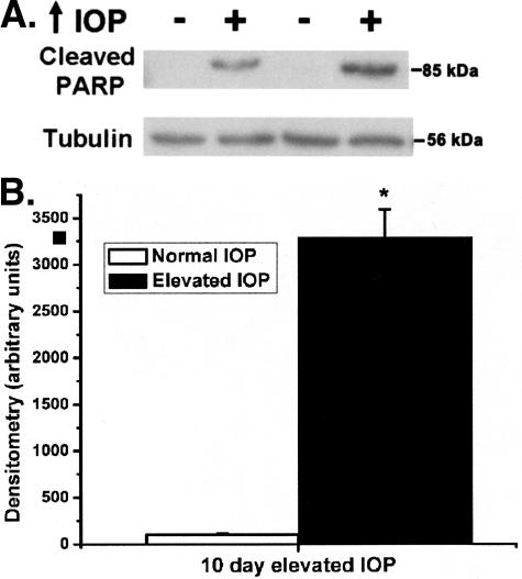
In glaucoma, retinal ganglion cells (RGCs) die by apoptosis, generally attributed to an elevated intraocular pressure (IOP). We now describe the impact of elevated IOP in the rat on expression of caspase 8 and caspase 9, initiators of the extrinsic and intrinsic caspase cascades, respectively. Activation of both caspases was demonstrated by the presence of cleaved forms of the caspases and the detection of cleaved Bid and PARP, downstream consequences of caspase activation. Surprisingly, the absolute level of procaspase 9 was also elevated after 10 days of increased IOP. To examine the cause of increased levels of the procaspase, we used laser capture microdissection to capture Fluorogold back-labeled RGCs and real-time polymerase chain reaction to measure mRNA changes of initiating caspases. The mRNA levels of both caspase 8 and caspase 9 were increased specifically in RGCs. These data suggest that elevated IOP activates a transcriptional up-regulation and activation of initiating caspases in RGCs and triggers apoptosis through both extrinsic and intrinsic caspase cascades.
Figures


Similar articles
-
Mechanisms of retinal ganglion cell injury and defense in glaucoma.Exp Eye Res. 2010 Jul;91(1):48-53. doi: 10.1016/j.exer.2010.04.002. Epub 2010 Apr 13. Exp Eye Res. 2010. PMID: 20394744 Free PMC article. Review.
-
Activation of caspase 9 in a rat model of experimental glaucoma.Curr Eye Res. 2002 Dec;25(6):389-95. doi: 10.1076/ceyr.25.6.389.14233. Curr Eye Res. 2002. PMID: 12789547
-
Caspase activation and amyloid precursor protein cleavage in rat ocular hypertension.Invest Ophthalmol Vis Sci. 2002 Apr;43(4):1077-87. Invest Ophthalmol Vis Sci. 2002. PMID: 11923249
-
Downregulation of Thy1 in retinal ganglion cells in experimental glaucoma.Curr Eye Res. 2006 Mar;31(3):265-71. doi: 10.1080/02713680500545671. Curr Eye Res. 2006. PMID: 16531284
-
Glaucoma: ocular Alzheimer's disease?Front Biosci. 2003 Sep 1;8:s1140-56. doi: 10.2741/1172. Front Biosci. 2003. PMID: 12957857 Review.
Cited by
-
Glaucoma-induced degeneration of retinal ganglion cells prevented by hypoxic preconditioning: a model of glaucoma tolerance.Mol Med. 2012 May 9;18(1):697-706. doi: 10.2119/molmed.2012.00050. Mol Med. 2012. PMID: 22396016 Free PMC article.
-
Heat shock protein 70 (HSP70) is critical for the photoreceptor stress response after retinal detachment via modulating anti-apoptotic Akt kinase.Am J Pathol. 2011 Mar;178(3):1080-91. doi: 10.1016/j.ajpath.2010.11.072. Am J Pathol. 2011. PMID: 21356360 Free PMC article.
-
Rgcs1, a dominant QTL that affects retinal ganglion cell death after optic nerve crush in mice.BMC Neurosci. 2008 Jul 31;9:74. doi: 10.1186/1471-2202-9-74. BMC Neurosci. 2008. PMID: 18671875 Free PMC article.
-
Mechanisms of retinal ganglion cell injury and defense in glaucoma.Exp Eye Res. 2010 Jul;91(1):48-53. doi: 10.1016/j.exer.2010.04.002. Epub 2010 Apr 13. Exp Eye Res. 2010. PMID: 20394744 Free PMC article. Review.
-
Intraocular Pressure Induced Retinal Changes Identified Using Synchrotron Infrared Microscopy.PLoS One. 2016 Oct 6;11(10):e0164035. doi: 10.1371/journal.pone.0164035. eCollection 2016. PLoS One. 2016. PMID: 27711151 Free PMC article.
References
-
- Kerrigan LA, Zack DJ, Quigley HA, Smith SD, Pease ME. TUNEL-positive ganglion cells in human primary open-angle glaucoma. Arch Ophthalmol. 1997;115:1031–1035. - PubMed
-
- Wax MB, Tezel G, Edward PD. Clinical and ocular histopathological findings in a patient with normal-pressure glaucoma. Arch Ophthalmol. 1998;116:993–1001. - PubMed
-
- Garcia-Valenzuela E, Shareef S, Walsh J, Sharma SC. Programmed cell death of retinal ganglion cells during experimental glaucoma. Exp Eye Res. 1995;61:33–44. - PubMed
-
- Hanninen VA, Pantcheva MB, Freeman EE, Poulin NR, Grosskreutz CL. Activation of caspase 9 in a rat model of experimental glaucoma. Curr Eye Res. 2002;25:389–395. - PubMed
-
- Quigley HA, Nickells RW, Kerrigan LA, Pease ME, Thibault DJ, Zack DJ. Retinal ganglion cell death in experimental glaucoma and after axotomy occurs by apoptosis. Invest Ophthalmol Vis Sci. 1995;36:774–786. - PubMed
Publication types
MeSH terms
Substances
Grants and funding
LinkOut - more resources
Full Text Sources
Other Literature Sources
Medical
