. 2005 Sep;167(3):887-99.
doi: 10.1016/S0002-9440(10)62060-3.

CXCR 3 activation promotes lymphocyte transendothelial migration across human hepatic endothelium under fluid flow

Affiliations

CXCR 3 activation promotes lymphocyte transendothelial migration across human hepatic endothelium under fluid flow

Stuart M Curbishley et al. Am J Pathol. 2005 Sep.

Abstract

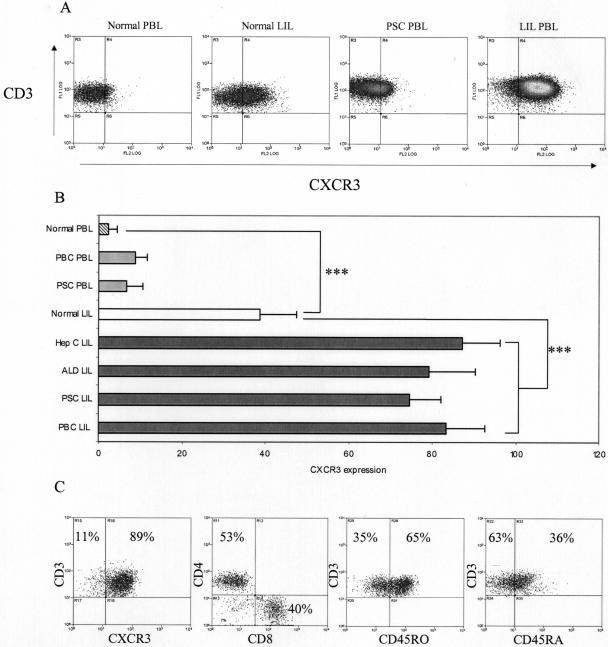
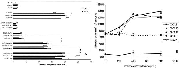
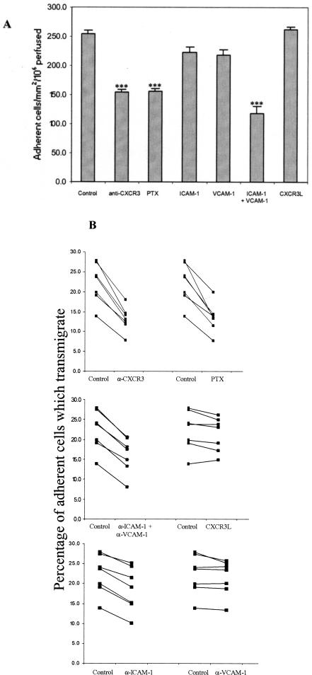
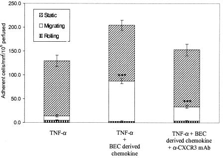
T cells infiltrating the inflamed liver express high levels of CXCR 3 and show enhanced migration to CXCR 3 ligands in chemotactic assays. Moreover, CXCR 3 ligands are up-regulated on hepatic endothelium at sites of T-cell infiltration in chronic hepatitis, and their presence correlates with outcome of inflammatory liver disease. We used a flow-based adhesion assay with human hepatic endothelium to investigate the function of CXCR 3 on lymphocyte adhesion to and transmigration through hepatic endothelium under physiological conditions of blood flow. To more accurately model the function of in vivo activated CXCR 3(high) lymphocytes, we isolated T cells from human liver tissue and studied their behavior in flow-based adhesion assays. We demonstrate that CXCR 3 not only promoted the adhesion of effector T cells to endothelium from flow but also drove transendothelial migration. Moreover, these responses could be stimulated either by endogenous CXCR 3 ligands secreted by the endothelium or by exogenous CXCR 3 ligands derived from other cell types and presented by the endothelium. This study thus demonstrates that activation of CXCR 3 promotes lymphocyte adhesion and transendothelial migration under flow and that human hepatic endothelium can present functionally active chemokines secreted by other cell types within the liver.


Figures

Figure 1
Figure 1
Expression of adhesion molecules determined by ELISA on HSECs (A) and BECs (B) in response to stimulation with a variety of cytokines. Data represent the mean ± SEM of four replicated experiments using different cell isolates. Values represent the mean absorbance of three replicate wells minus the absorbance of an isotype-matched control antibody. All cytokine treatments were at 10 ng/ml for 24 hours before assay. U/S, unstimulated; TNF-α/β, tumor necrosis factor-α/β; IL-1β, interleukin-1β; OSM, oncostatin M.
Figure 2
Figure 2
Secretion of CXCR3 ligands by HSECs (A) and BECs (B) in response to stimulation with a variety of cytokines. Data represent the mean ± SEM of four replicate capture ELISA experiments using different cell isolates. Values represent the mean absorbance of three replicate wells minus the absorbance of a blank well. All cytokine treatments were at 10 ng/ml for 24 hours before assay. No detectable chemokine was secreted by cytokine-stimulated cells in the absence of IFN-γ treatment.
Figure 3
Figure 3
De novo synthesis of CXCR3 ligands by human sinusoidal endothelial cells was confirmed by RT-PCR. In the absence of any stimulus no product was detected. Stimulation with TNF-α had little or no effect on CXCL9/11 production but did up-regulate CXCL10. Stimulation with TNF-α concomitant with IFN-γ consistently up-regulated all three CXCR3L. Representative data from one of three replicate experiments are shown.
Figure 4
Figure 4
Expression of CXCR3 ligands in human liver samples. I: Top: Low-power photomicrographs demonstrating expression (brown pigment) of CXCL9 (A), CXCL10 (B), and CXCL11 (C) in chronically inflamed human liver (primary biliary cirrhosis) tissue by immunohistochemistry. All three ligands were detected on sinusoids with particularly strong staining at areas of inflammation and active fibrosis (A, arrow). Little or no expression was detected in normal liver tissue (D–F). I: Bottom: High-power photomicrographs demonstrating CXCL11 expression on hepatocyte membranes (G) and CXCL10 expression on liver sinusoids (H). J: CXCL11 expression was also detected on bile ducts and proliferating bile ductules. Staining with an isotype-matched control antibody was negative for all samples. II: Western blot analysis of whole liver tissue lysates (B) was used to confirm expression. Marked levels of CXCL9, -10, and -11 were recorded in viral as well as chronically inflamed liver samples whereas little chemokine was detected in normal liver samples. PSC, primary sclerosing cholangitis; PBC, primary biliary cirrhosis; Hep C, chronic hepatitis C infection; ALD, alcoholic liver disease; NL, normal liver. Original magnifications: ×200 (I); ×400 (II).
Figure 5
Figure 5
Little CXCR3 was detected on peripheral blood lymphocytes (PBLs) from normal patients with a small but significant increase on peripheral blood from patients with chronic inflammatory liver diseases. The number of cells expressing high levels of CXCR3 was significantly increased on LILs (P < 0.001) and further increased on LILs isolated from diseased livers (P < 0.001). A: Representative dot plots of CXCR3- and CD3-labeled LILs from normal and diseased livers. B: The percentage of cells that expressed CXCR3 on lymphocytes isolated from normal liver and chronically inflamed liver is derived from at least three experiments for each group shown as mean ± SEM. C: Phenotypic analysis of liver-infiltrating T lymphocytes (CD3+) revealed that on average 89% were CXCR3+, compared to just 4% in peripheral blood. The ratio of CD4- to CD8-positive lymphocytes on CXCR3+ T cells was 55:45, with 65% displaying classical memory phenotype (CD45RO+). The 35% CD45RA+ cells probably represent terminally differentiated effector cells that have re-expressed CD45RA rather than true naïve cells. Representative data from one of three replicate experiments. PSC, primary sclerosing cholangitis; PBC, primary biliary cirrhosis; Hep C, chronic hepatitis C infection; ALD, alcoholic liver disease.
Figure 6
Figure 6
CXCR3 ligands trigger integrin-mediated adhesion of LILs to ICAM-1 and VCAM-1 under static conditions (A) and to VCAM-1 under flow (B). A: Lymphocytes isolated from chronically inflamed liver samples or matched peripheral blood samples were resuspended to a count of 8 × 10⁴ ml and applied to individual wells of an 18-well slide, precoated with ICAM-1 (5 μg/ml). Co-incubation with CXCL9, -10, or -11 significantly increased the binding of LILs, although this effect was abrogated by the addition of pertussis toxin (PTX). Co-incubation of peripheral blood lymphocytes (PBLs) with CXCR3 ligands did not trigger adhesion. Adhesion of LILs and peripheral blood lymphocytes was increased in the presence of the integrin activator MnCl2. Data represent mean ± SEM for five replicate experiments. Statistical significance was calculated using paired t-tests, comparing lymphocytes isolated from diseased or normal livers (grouped) with those from the peripheral circulation. B: Adhesion of LILs to immobilized VCAM-1 and ICAM-1 under flow. VCAM-1 or ICAM-1 was immobilized (5 μg/ml) onto glass microslides and adhesion triggered by co-immobilization of the chemokines CXCL9, -10, and -11. CXCL5 was used to control for any nonspecific effects of chemokine co-immobilization because very few lymphocytes express its receptor CXCR2. No adhesion from flow was detected with ICAM-1, even in the presence of CXCR3 ligands. VCAM-1 was able to capture lymphocytes efficiently from flow and co-immobilization of CXCR3 ligands enhanced this effect significantly adhesion being maximal at a chemokine concentration of 400 ng/ml (P < 0.001). Data represent mean ± SEM for three replicate experiments performed at 0.05 Pa.
Figure 7
Figure 7
CXCR3 ligands promote lymphocyte adhesion from flow and subsequent transmigration on TNF-α/IFN-γ-stimulated human HSECs. A: Inhibition of CXCR3 with a function blocking antibody or a Gi protein block (PTX) significantly reduces total adhesion of LILs from flow to TNF-α/IFN-γ-stimulated HSECs. Adhesion was further reduced by the addition of blocking antibodies against ICAM-1/VCAM-1. Data represent number of adherent cells per mm2 per million perfused and are the mean ± SEM of seven experiments. B: Transmigration of adherent lymphocytes through TNF-α/IFN-γ-stimulated HSECs was significantly reduced after CXCR3 and ICAM-1 blockade whereas anti-VCAM-1 mAb had no affect either when used alone or when used in combination. Migrating cells were calculated using frame-by-frame analysis of experimental videos to count the percentage of phase dark (transmigrated) lymphocytes. Data represent paired samples from individual experiments. All experiments were performed at 0.05 Pa.
Figure 8
Figure 8
Incubation of TNF-α-stimulated HSECs with BEC-derived CXCR3 ligands increased total adhesion and transendothelial migration of liver-derived lymphocytes under conditions of flow. Pretreatment of HSEC monolayers with BEC supernatant containing CXCL9, -10, and -11 increased the number of adherent lymphocytes. No variation in the number of static nonmigrating lymphocytes was recorded but the number of lymphocytes transmigrating was significantly increased (P = 0.001). Pretreatment of lymphocytes with a CXCR3 blocking antibody significantly reduced the number of lymphocytes migrating across the HSECs. Data represent the mean of three replicate experiments ± SEM. All experiments were performed at 0.05 Pa.

References

    1. Campbell JJ, Butcher EC. Chemokines in tissue-specific and microenvironment-specific lymphocyte homing. Curr Opin Immunol. 2000;12:336–341. - PubMed
    1. von Andrian UH, Mackay CR. T-cell function and migration. Two sides of the same coin. N Engl J Med. 2000;343:1020–1034. - PubMed
    1. Pachynski RK, Wu SW, Gunn MD, Erle DJ. Secondary lymphoid-tissue chemokine (SLC) stimulates integrin alpha 4 beta 7-mediated adhesion of lymphocytes to mucosal addressin cell adhesion molecule-1 (MAdCAM-1) under flow. J Immunol. 1998;161:952–956. - PubMed
    1. Foxman EF, Kunkel EJ, Butcher EC. Integrating conflicting chemotactic signals. The role of memory in leukocyte navigation. J Cell Biol. 1999;147:577–588. - PMC - PubMed
    1. Foxman EF, Campbell JJ, Butcher EC. Multistep navigation and the combinatorial control of leukocyte chemotaxis. J Cell Biol. 1997;139:1349–1360. - PMC - PubMed

Publication types

MeSH terms

LinkOut - more resources