. 2005 Sep;115(9):2434-43.
doi: 10.1172/JCI24819. Epub 2005 Aug 25.

Human aldose reductase expression accelerates diabetic atherosclerosis in transgenic mice

Affiliations

Human aldose reductase expression accelerates diabetic atherosclerosis in transgenic mice

Reeba K Vikramadithyan et al. J Clin Invest. 2005 Sep.

Abstract

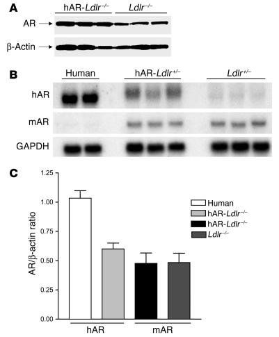
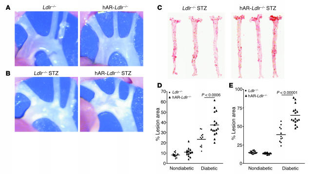
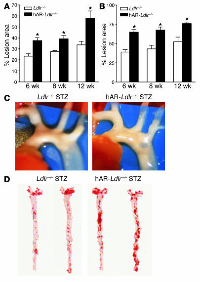
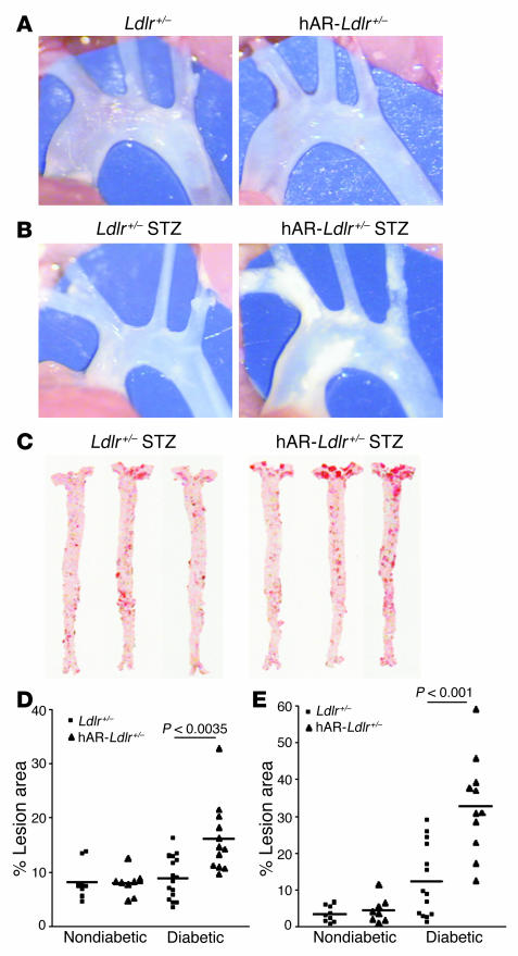
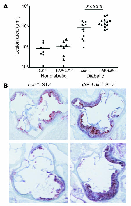
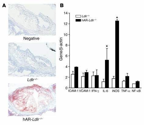
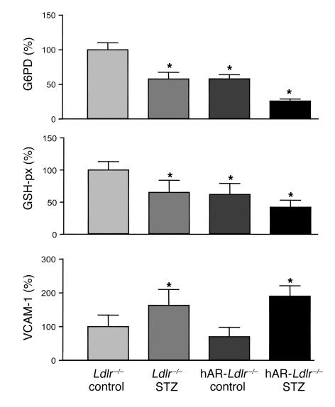
Direct evidence that hyperglycemia, rather than concomitant increases in known risk factors, induces atherosclerosis is lacking. Most diabetic mice do not exhibit a higher degree of atherosclerosis unless the development of diabetes is associated with more severe hyperlipidemia. We hypothesized that normal mice were deficient in a gene that accelerated atherosclerosis with diabetes. The gene encoding aldose reductase (AR), an enzyme that mediates the generation of toxic products from glucose, is expressed at low levels in murine compared with human tissues. Mice in which diabetes was induced through streptozotocin (STZ) treatment, but not nondiabetic mice, expressing human AR (hAR) crossed with LDL receptor-deficient (Ldlr-/-) C57BL/6 male mice had increased aortic atherosclerosis. Diabetic hAR-expressing heterozygous LDL receptor-knockout mice (Ldlr+/-) fed a cholesterol/cholic acid-containing diet also had increased aortic lesion size. Lesion area at the aortic root was increased by STZ treatment alone but was further increased by hAR expression. Macrophages from hAR-transgenic mice expressed more scavenger receptors and had greater accumulation of modified lipoproteins than macrophages from nontransgenic mice. Expression of genes that regulate regeneration of glutathione was reduced in the hAR-expressing aortas. Thus, hAR increases atherosclerosis in diabetic mice. Inhibitors of AR or other enzymes that mediate glucose toxicity could be useful in the treatment of diabetic atherosclerosis.


Figures

Figure 1
Figure 1
Expression of AR in transgenic mice, normal human hearts, and macrophages. (A) Western blot of AR protein in aortas from Ldlr–/– and hAR-Ldlr–/– mice. Aortas were obtained from 8-week-old mice on chow diets, the aortas were harvested and homogenized, and equal amounts of protein were loaded on SDS gels. AR was detected using a polyclonal antibody that recognizes both human and mAR. (B) Northern blot analysis of heart expression of human and mAR gene. (C) Real-time PCR analysis of macrophage expression of hAR and mAR mRNA. Values are expressed as mean ± SEM; n = 5 mice per group. For human macrophages, 2 samples were assayed in duplicate 3 times.
Figure 2
Figure 2
Diabetes increased the atherosclerosis lesion size in male hAR-Ldlr–/– mice fed the HCD for 6 weeks. (A) Aortic arch lesion area in Ldlr–/– and hAR-Ldlr–/– nondiabetic mice. (B) Aortic arch lesion area in Ldlr–/– and hAR-Ldlr–/– diabetic mice. Magnification, ×26. (C) Oil red O staining of en face lesion area of Ldlr–/– and hAR-Ldlr–/– diabetic mice. (D) Total lesion area in nondiabetic and diabetic Ldlr–/– and hAR-Ldlr–/– mice. (E) Aortic arch lesion area in nondiabetic and diabetic Ldlr–/– and hAR-Ldlr–/– mice.
Figure 3
Figure 3
The atherosclerosis lesion size increased in diabetic mice with increased time period of high-cholesterol feeding. (A) Total lesion area in diabetic Ldlr–/– and hAR-Ldlr–/– mice after 6, 8, and 12 weeks. (B) Lesion in the aortic arch of diabetic Ldlr–/– and hAR-Ldlr–/– mice after 6, 8, and 12 weeks on the HCD. Values are mean ± SEM of 5–8 mice per group; *P < 0.05. (C) Aortic arch lesion area in 12-week HCD-fed Ldlr–/– and hAR-Ldlr–/– diabetic mice. Magnification, ×26. (D) Oil red O staining of en face lesion area of Ldlr–/– and hAR-Ldlr–/– diabetic mice fed the HCD for 12 weeks.
Figure 4
Figure 4
Diabetic hAR-Ldlr+/– mice have greater lesion area than Ldlr+/– mice fed a CCA diet for 12 weeks. (A) Aortic arch lesion area in Ldlr+/– and hAR-Ldlr+/– nondiabetic mice. (B) Aortic arch lesion area in Ldlr+/– and hAR-Ldlr+/– diabetic mice. Magnification, ×26. (C) Oil red O staining of en face lesion area in Ldlr+/– and hAR-Ldlr+/– diabetic mice. (D) Total lesion area in nondiabetic and diabetic Ldlr+/– and hAR-Ldlr+/– mice (E) Aortic arch lesion area in nondiabetic and diabetic Ldlr+/– and hAR-Ldlr+/– mice.
Figure 5
Figure 5
Diabetes increased the atherosclerosis lesion size in the aortic root area of hAR-Ldlr–/– mice fed the CCA diet for 12 weeks. (A) Aortic root atherosclerotic lesion area in Ldlr+/– and hAR-Ldlr+/– mice. (B) Oil red O–stained sections of the root area of diabetic Ldlr+/– and hAR-Ldlr+/– mice. Two representative mice from each group are shown. Magnification, ×100.
Figure 6
Figure 6
hAR-expressing mice have more AR protein expression in the atherosclerotic lesion area compared with nontransgenic mice. iNOS and IL-6 increased several folds in peritoneal macrophages from hAR-Ldlr–/– compared with those from Ldlr–/– mice. (A) Immunohistochemical staining of aortic root sections of Ldlr–/– and hAR-Ldlr–/– diabetic mice for AR protein using hAR antibody. “Negative” indicates staining after use of nonimmune serum. Magnification, ×200. (B) Real-time PCR gene expression of iNOS, IL-6, ICAM-1, VCAM-1, IFN-γ, TNF-α, and NF-κB in peritoneal macrophages from Ldlr–/– and hAR-Ldlr–/– mice. Values were normalized to β-actin and expressed as mean ± SEM. Data are from experiments performed in duplicate on an average of 8 mice. *P < 0.05.
Figure 7
Figure 7
Expression of modified lipoprotein receptors and uptake of Ac-LDL are increased in peritoneal macrophages. (A) mRNA expression of CD36 and SR-A by real-time PCR in peritoneal macrophages from Ldlr–/– and hAR-Ldlr–/– mice. Values were normalized to β-actin and expressed as mean ± SEM. Data are from experiments performed in duplicate on an average of 8 mice. (B) Western blot analysis of CD36, SR-A, and ATP-binding cassette A1 (ABCA1) protein in peritoneal macrophages from hAR-Ldlr–/– and Ldlr–/– mice. Data are from pooled macrophages collected from 3 mice per group. (C) Uptake of 125I-Ac-LDL (100 μg/ml) in peritoneal macrophages from Ldlr–/– and hAR-Ldlr–/– mice. Values are expressed as mean ± SEM. Data are from experiments performed in duplicate on 6 mice. *P < 0.05.
Figure 8
Figure 8
Aortic gene changes prior to atherosclerosis. Aortas were obtained from Ldlr–/– and hAR-Ldlr–/– mice either left untreated (control) or made diabetic by STZ injection. There were 4–6 aortas in each of the 4 groups. The STZ treatment occurred 4 weeks earlier, and the mice remained on a chow diet. Gene expression was assessed by real-time PCR. *P < 0.05 versus control. G6PD and GSH-px mRNA levels in STZ-treated hAR-Ldlr–/– aortas were also significantly reduced (P < 0.05) compared with those in STZ-treated Ldlr–/– aortas.

References

    1. Anonymous Intensive blood-glucose control with sulphonylureas or insulin compared with conventional treatment and risk of complications in patients with type 2 diabetes (UKPDS 33). UK Prospective Diabetes Study (UKPDS) Group. Lancet. 1998;352:837–853. - PubMed
    1. Diabetes ControlComplications Trial. Effect of intensive diabetes management on macrovascular events and risk factors in the Diabetes Control and Complications Trial. Am. J. Cardiol. 1995;75:894–903. - PubMed
    1. Goldberg IJ. Why does diabetes increase atherosclerosis? I don’t know! J. Clin. Invest. 2004;114:613–615. doi:10.1172/JCI200422826. - PMC - PubMed
    1. Mathe D. Dyslipidemia and diabetes: animal models [review] Diabete Metab. 1995;21:106–111. - PubMed
    1. Keren P, et al. Effect of hyperglycemia and hyperlipidemia on atherosclerosis in LDL receptor-deficient mice: establishment of a combined model and association with heat shock protein 65 immunity. Diabetes. 2000;49:1064–1069. - PubMed

Publication types

MeSH terms