. 2005 Sep;115(9):2373-81.
doi: 10.1172/JCI25118. Epub 2005 Aug 25.

Activating and deactivating mutations in the receptor interaction site of GDF5 cause symphalangism or brachydactyly type A2

Affiliations

Activating and deactivating mutations in the receptor interaction site of GDF5 cause symphalangism or brachydactyly type A2

Petra Seemann et al. J Clin Invest. 2005 Sep.

Abstract

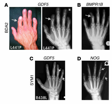
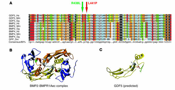
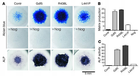
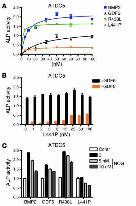
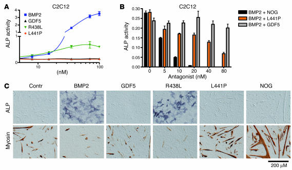
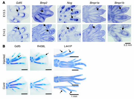
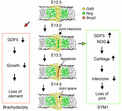
Here we describe 2 mutations in growth and differentiation factor 5 (GDF5) that alter receptor-binding affinities. They cause brachydactyly type A2 (L441P) and symphalangism (R438L), conditions previously associated with mutations in the GDF5 receptor bone morphogenetic protein receptor type 1b (BMPR1B) and the BMP antagonist NOGGIN, respectively. We expressed the mutant proteins in limb bud micromass culture and treated ATDC5 and C2C12 cells with recombinant GDF5. Our results indicated that the L441P mutant is almost inactive. The R438L mutant, in contrast, showed increased biological activity when compared with WT GDF5. Biosensor interaction analyses revealed loss of binding to BMPR1A and BMPR1B ectodomains for the L441P mutant, whereas the R438L mutant showed normal binding to BMPR1B but increased binding to BMPR1A, the receptor normally activated by BMP2. The binding to NOGGIN was normal for both mutants. Thus, the brachydactyly type A2 phenotype (L441P) is caused by inhibition of the ligand-receptor interaction, whereas the symphalangism phenotype (R438L) is caused by a loss of receptor-binding specificity, resulting in a gain of function by the acquisition of BMP2-like properties. The presented experiments have identified some of the main determinants of GDF5 receptor-binding specificity in vivo and open new prospects for generating antagonists and superagonists of GDF5.


Figures

Figure 1
Figure 1
Phenotypes of the right hands of patients carrying L441P and R438L mutations. (A) The L441P mutation is associated with brachydactyly, characterized by a short index finger and bending of finger V (clinodactyly). X-rays show missing middle phalanges in finger II and hypoplasia of the middle phalanges in finger V. (B) The phenotype is very similar to that of BDA2, which is caused by mutations in BMPR1B. (C) The R438L mutation results in SYM1, characterized by bony fusion of the proximal interphalangeal joint of finger V and an abnormal interphalangeal joint in finger IV. (D) The phenotype is very similar to that of SYM1, which is caused by mutations in NOG.
Figure 2
Figure 2
Protein sequence alignment of GDF5 homologs and 3D models for GDF5 receptor binding. (A) Primary sequence alignment of GDF5, BMP2, and BMP4 from different species, including drosophila DPP. The positions of R438L and L441P mutations in GDF5 are indicated by arrows. Note the widespread conservation of residue L441 in BMPs and the specificity of R438 conservation for the GDF5 subfamily (replaced by A in the BMPs). (B) 3D model of a BMP2 dimer (yellow and gold) linked via a disulfide bridge and bound to the ectodomain of BMPR1A (BMPR1Aec). The amino acids mutated in GDF5 are indicated in red (L441) and green (R438). Note their position within the receptor interaction site. (C) Predicted structure of a GDF5 monomer. The positions of the mutated amino acids are indicated in red (L441) and green (R438).
Figure 3
Figure 3
Functional analysis of Gdf5 mutants in micromass culture. (A) Chicken micromass cultures were assayed after 4 days for extracellular matrix production and analyzed after 7 days for ALP activity. Cells were infected with virus containing WT Gdf5 or mutant sequences and coinfected with Nog or not coinfected. The mutants differed drastically in their biological activity. Coinfection with Nog completely represses chondrogenesis irrespective of the Gdf5 variant expressed. Magnification, ×1 objective (AxioCam HRc camera; Zeiss). (B) Alcian blue incorporation into the extracellular matrix of micromass cultures reflecting the production of proteoglycan-rich cartilaginous matrix measured at day 4 was quantified after extraction. Biological activity of R438L mutant was comparable to that of Gdf5, whereas the L441P mutant displayed only small effects on matrix production. (C) ALP activity of micromass cultures at day 7 was quantified by a specific enzymatic assay. While Gdf5 and R438L induced ALP activity effectively, L441P caused only a small amount of induction, slightly above control activity. Contr, control.
Figure 4
Figure 4
Characterization of ALP induction by GDF5 mutants in chondrogenic ATDC5 cells. ALP activity was measured after stimulation of ATDC5 cells with increasing amounts of recombinant proteins after 3 days. (A) Recombinant BMP2, GDF5, and R438L display characteristic dose-dependent inductions of ALP activity in ATDC5 cells. The R438L mutant induces significantly higher ALP activity than WT GDF5, whereas L441P is almost inactive. (B) Stimulation of ATDC5 cells with L441P or a combination of GDF5 and L441P. Cells were incubated with increasing amounts of L441P with or without 10 nM GDF5. No stimulating or suppressing effect of increasing amounts of L441P on GDF5-dependent induction of ALP activity was observed. (C) Inhibition of ALP induction by recombinant NOG in stimulated ATDC5 cells. ALP was determined after incubation with 5 nM recombinant BMP2, GDF5, R438L, or L441P, and cotreatment with increasing amounts of NOG. GDF5, R438L, and L441P display similar inhibition profiles by NOG.
Figure 5
Figure 5
Effects of GDF5 mutants on the differentiation of premyoblastic C2C12 cells. ALP activity was analyzed after stimulation of C2C12 cells with the recombinant proteins for 3 days. Differentiation markers ALP and myosin were assayed 5 days after addition of the proteins. (A) WT GDF5 and the L441P mutant did not induce significant ALP activity in C2C12 cells. In contrast, treatment with BMP2 resulted in a strong induction, and the R438L mutant induced significant ALP activity, albeit to a lower degree than BMP2. (B) Addition of NOG inhibited BMP2-induced ALP activity in a dose-dependent fashion. A similar antagonism was observed with the addition of L441P mutant. The addition of WT GDF5 had no major effect. (C) Differentiation of C2C12 cells was determined using ALP staining (osteoblastic lineage) or immunohistochemical analysis of myosin expression (muscle lineage). Treatment of C2C12 cells with WT GDF5 had a slightly negative effect on their spontaneous differentiation along the muscle lineage, as indicated by the reduced size of myoblasts. L441P treatment increased muscle formation with results similar to the effects of the BMP inhibitor NOG. Incubation with BMP2, in contrast, resulted in strong induction of ALP activity and suppression of muscle differentiation. Similar results were obtained for the R438L mutant, indicating that this mutant displayed BMP2-like activity in this assay. Magnification, ×10 objective (AxioCam HRc camera; Zeiss).
Figure 6
Figure 6
Expression analysis during joint development and overexpression of Gdf5 in vivo. (A) In situ hybridization on mouse limb sections at E13.5 and E14.5 with probes specific for Gdf5, Bmp2, Nog, Bmpr1a, and Bmpr1b. The area of joint formation is indicated by arrows. Note strong expression of Gdf5 at both stages but Bmp2 expression in the joint only at E14.5. The longitudinal stripes of Bmp2 expression in the joint area at E13.5 correspond to expression in developing ligaments and not the joint interzone. Nog was expressed in a small band of cells in the joint interzone at E13.5 but not at E14.5. Bmpr1b was expressed in chondrocytes flanking the joint interzone. Bmpr1a shows ubiquitous expression with higher levels in the perichondrium and the developing joints. (B) Overexpression of WT Gdf5 as well as the Gdf5 mutants R438L and L441P in chick embryos using RCAS retroviral system. Alcian blue staining was used to visualize cartilage. For comparison, the injected left limb is shown next to the uninfected right limb, which serves as the control. Note enlargement of skeletal elements, joint fusions, and fusions between digits in WT Gdf5 and R438L mutant infected limbs at stage HH32. Changes in the L441P-injected limb were less severe and were only observed at a later stage (HH34–35). A higher magnification (arrows in B) shows joint fusions. Magnification, ×10 objective (A); ×1.6 objective (WT Gdf5 and R438L in B); ×1.25 objective (L441P in B) (AxioCam HRc camera; Zeiss).
Figure 7
Figure 7
Schematic drawing of proposed mechanism of metacarpophalangeal joint development and expression of Gdf5, Nog, and Bmp2 in mouse limbs corresponding to E12.5, E13.0, E13.5, and E14.5. Gdf5 induces longitudinal growth at the distal end of each element. Bmp inhibitor Nog is expressed in the cartilage anlagen and the joint interzone first, while Bmp2 is expressed later. Apoptosis, mechanical forces, and other factors lead to formation of a joint space. Boxes on left and right show proposed molecular pathogenesis for brachydactyly and SYM1, respectively. Reduction of GDF5 signal, as in the L441P mutant, results in reduced growth of phalanges and thus shortening of elements. Increased GDF5 signaling, as in the R438L mutant, or mutations in NOG, both result in the persistence of cartilage in the joint region and a deficiency in joint interzone formation. Clinically this results in lack of joint formation and thus SYM1.

References

    1. Kornak U, Mundlos S. Genetic disorders of the skeleton: a developmental approach [review] Am. J. Hum. Genet. 2003;73:447–474. - PMC - PubMed
    1. Sebald W, Nickel J, Zhang JL, Mueller TD. Molecular recognition in bone morphogenetic protein (BMP)/receptor interaction. Biol. Chem. 2004;385:697–710. - PubMed
    1. Luyten FP. Cartilage-derived morphogenetic protein-1. Int. J. Biochem. Cell Biol. 1997;29:1241–1244. - PubMed
    1. Nishitoh H, et al. Identification of type I and type II serine/threonine kinase receptors for growth/differentiation factor-5. J. Biol. Chem. 1996;271:21345–21352. - PubMed
    1. Kirsch T, Nickel J, Sebald W. BMP-2 antagonists emerge from alterations in the low-affinity binding epitope for receptor BMPR-II. EMBO J. 2000;19:3314–3324. - PMC - PubMed

Publication types

MeSH terms