. 2005 Oct 17;491(2):109-22.
doi: 10.1002/cne.20714.

Contributions of cortical subventricular zone to the development of the human cerebral cortex

Affiliations

Contributions of cortical subventricular zone to the development of the human cerebral cortex

Nada Zecevic et al. J Comp Neurol. .

Abstract

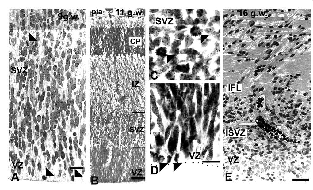
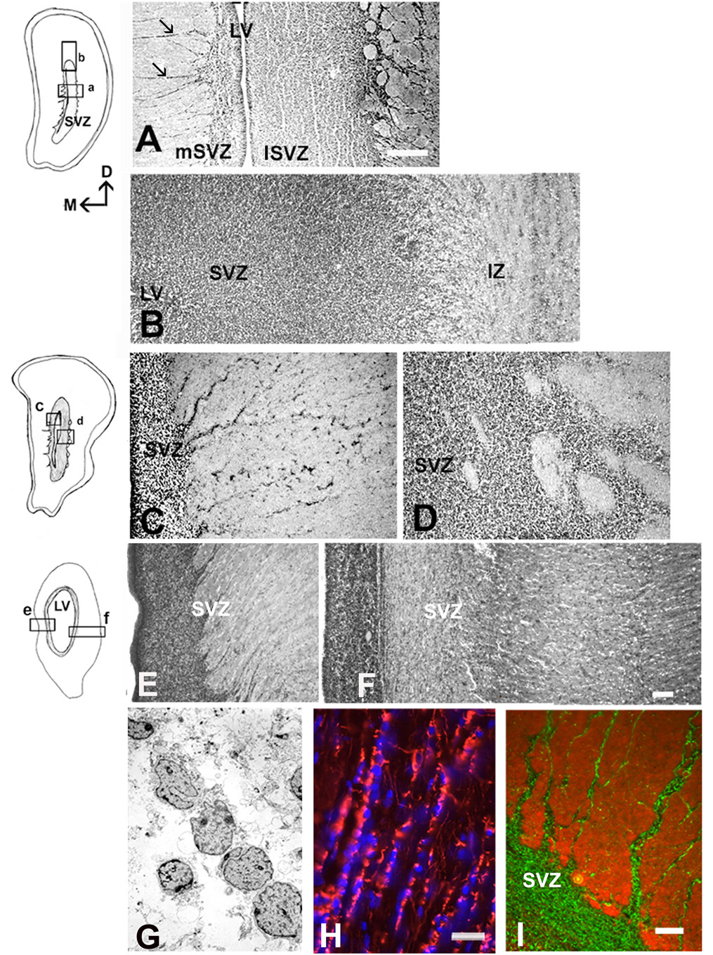
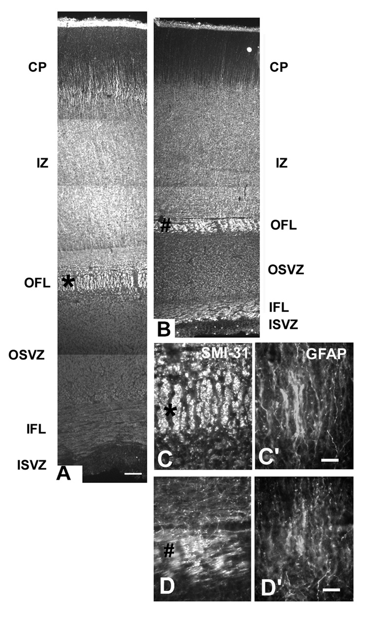
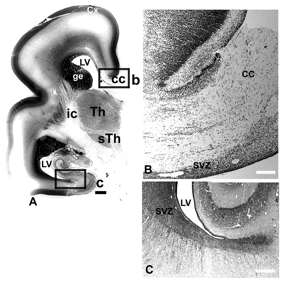
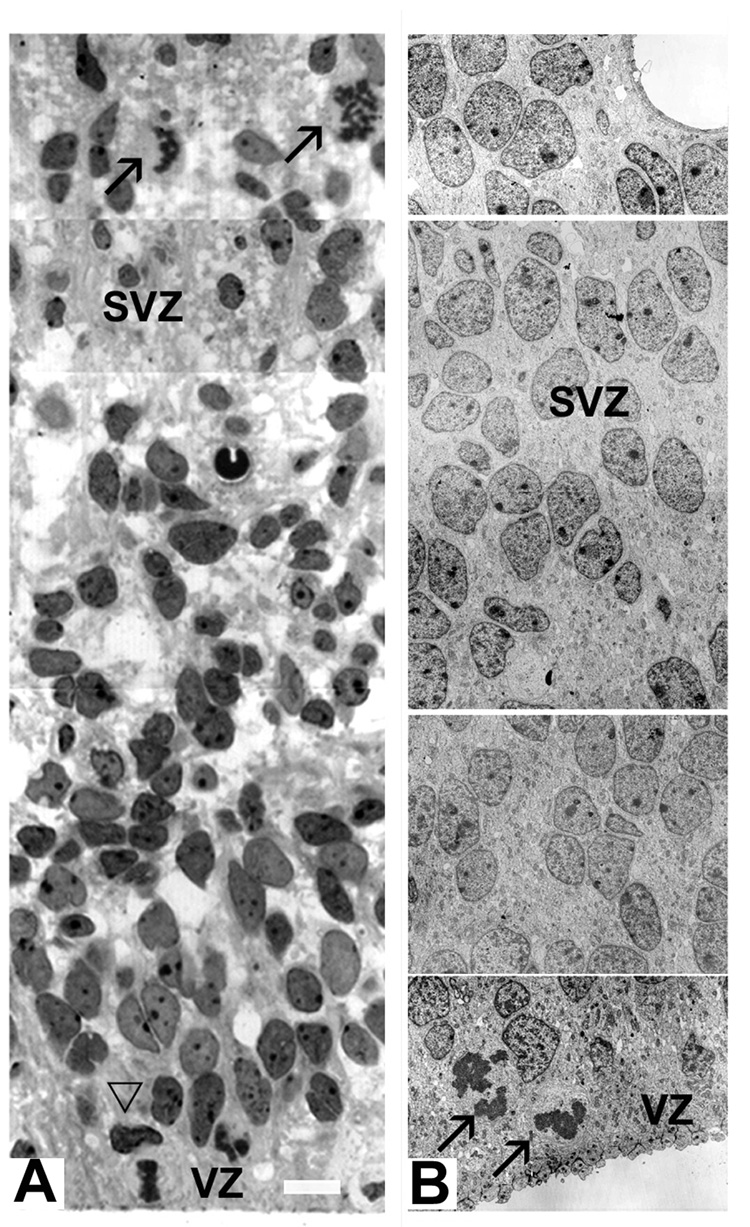
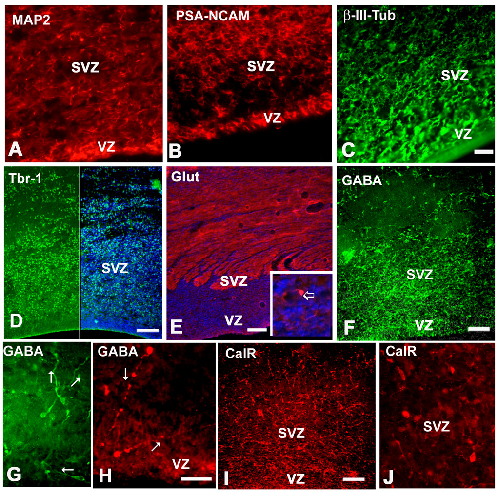
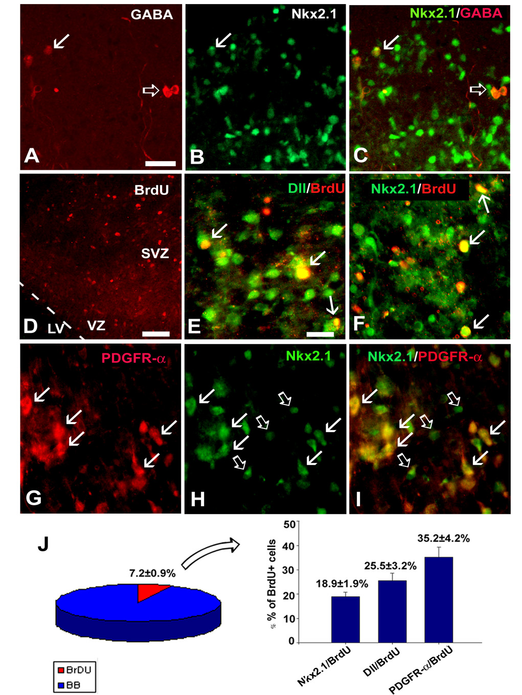
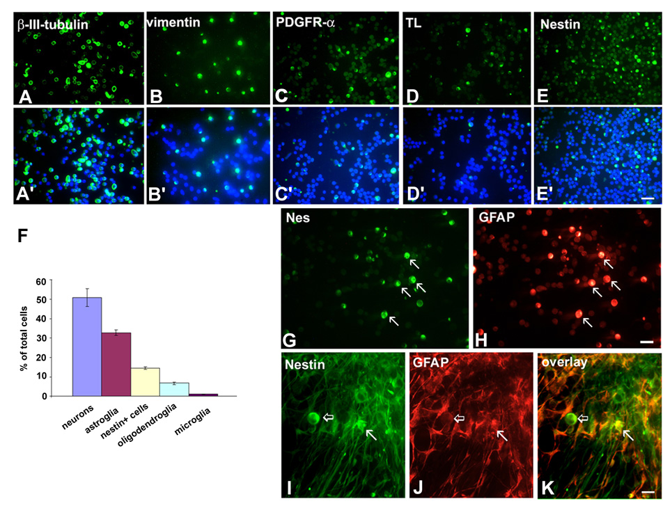
The cortical subventricular zone (SVZ), a proliferative compartment in the forebrain, has a uniquely important role during the second half of intrauterine development in human. This is best observed in numerous neonatal pathologies that result from prenatal SVZ damage. These conditions highlight a need to better understand the contribution of the SVZ to the development of the human cerebral cortex. With this goal in mind, we analyzed histological organization, cell proliferation, and molecular diversity in the human fetal SVZ from 7-27 gestational weeks (gw) using light and electron microscopy, immunohistochemistry, and in vitro methods. Complex histological organization distinguishes human cortical SVZ from that of other mammals. In vitro quantification showed that approximately 50% of cells in the VZ/SVZ region are neurons, 30% are astroglia, 15% are nestin+ cells, with other cell types representing smaller fractions. Immunolabeling with BrdU showed that a considerable number of cells ( approximately 10%) are generated in the human cortical SVZ during midgestation (18-24 gw) under in vitro conditions. Immunofluorescence with cell type-specific markers and BrdU revealed that all major cell types, neural precursors (nestin+), astroglia including radial glia (GFAP+, vimentin+), and oligodendrocyte progenitors (PDGFR-alpha+) were proliferating. An increase in the ratio of the size of the SVZ to VZ, protracted period of cell proliferation, as well as cellular and histological complexity of the human fetal SVZ are directly related to the evolutionary expansion of the human cerebral cortex.


Figures

Fig. 1
Fig. 1
VZ/SVZ region in the fetal human brain as viewed by light microscopy. A: At 9 gw Nissl stained section, the VZ merges with the SVZ without a sharp border; dividing cells (arrowheads) can be observed in both the VZ and SVZ. B: At 11 gw, in the lateral cortex, the SVZ/VZ border as well as the upper border with the intermediate zone (IZ) are clearly pronounced (lines on the right); C and D: Higher magnification of proliferating cells at 11 gw in C: the SVZ, and D: the VZ (arrowheads). E: In the occipital cortex at 17 gw, afferent fibers in the SVZ form the inner fiber layer (IFL). Scale bars= A, C, D -10µm, B-50µm, E-100µm.
Fig. 2
Fig. 2
Changing appearance of the SVZ along the rostro-caudal axis in the fetal brain at midgestation (22 gw). Drawings represent Nissl stained coronal sections; small quadrants mark areas represented on the photographs. A: In the frontal pole, the lateral SVZ (lSVZ) is five times wider than the medial SVZ (mSVZ) facing the interhemispheric fissure. Note the cell bands on the medial side (arrows). B: Dorsal SVZ from the same section. VZ is to the left, pia to the right. Fiber bundles are running at the border of the SVZ with the intermediate zone (IZ). C, D: More caudally, at the level of the internal capsule (IC), medial (C) and lateral SVZ (D). E, F: At the occipital pole level, the medial SVZ (E) with cell bands that stretch to the subplate layer; the lateral SVZ (F) has complex organization (see Fig.3 for details). G: On the electron micrograph of the SVZ at 22 gw, the cells in bands are closely linked. H: Cell nuclei counterstained with bis-benzimide (blue) aligned along GFAP+ fibers (red) radiating from the SVZ. I: Double-labeling with GFAP (green) and PSA-NCAM (red) reveals that these two markers do not overlap in the SVZ cell bands. Scale bars= A-500µm, B–F (in F)-100µm; G-Direct Mg.2000X, H-20µm, I- 50µm.
Fig. 3
Fig. 3
Occipital cortex at midgestation immunostained with SMI-31 antibody to display inner fiber layer (IFL) close to the inner SVZ, and the outer fiber layer (OFL) between the IZ and the outer SVZ (OSVZ). A: lateral and B: medial side of the hemisphere. A much narrower medial cerebral cortex demonstrates a smaller SVZ. IZ- the intermediate zone, CP-cortical plate. C-C”: Higher magnification of the outer fiber layer in the lateral SVZ labeled with an asterisk on A. Double labeling with SMI-31 (C) and GFAP (C’) reveals characteristic “palisades” of radially aligned SMI-31+ cells and GFAP+ fibers. D,D’: In the medial SVZ, in contrast, this palisade organization is not present; D: SMI-31, and D’: GFAP. Scale bars= A, B-100µm; C,D- 10µm.
Fig. 4
Fig. 4
Nissl stained frontal section through the mid-brain level at 25gw. A: Contact picture with boxed areas which are represented on corresponding photographs. B: Medial SVZ in the intrahemispheric fissure, with cell bands extending into the corpus callosum (cc). C: Posterior SVZ with cell stream invading the white matter of the temporal pole. SVZ-subventricular zone, Th-thalamus, sTh- subthalamic nucleus, LV-lateral ventricle, ge-ganglionic eminence, cc-corpus callosum, ic-internal capsule, Cx-cerebral cortex. . Scale bars= A-1mm, B, C-100µm.
Fig. 5
Fig. 5
Various shape and staining properties of SVZ cells on the light and electron microscopic level. A: On a 1µm toluidine blue stained section of the 18 gw fetus, diverse staining properties of SVZ cells could be seen, along with horizontal and vertical mitotic spindles on the VZ surface (empty arrowhead) and in the SVZ (arrows). B: On the electron micrograph at 12 gw darker and lighter cell nuclei are dispersed through the SVZ, whereas two mitoses are observed at the ventral surface (arrows). Scale bars= A-10µm; B - direct Mg. 2000X.
Fig. 6
Fig. 6
Immunohistochemical labeling of different cortical neuron subtypes in the SVZ at midgestation. A: MAP2, B: PSA-NCAM, C: β-III-tubulin, D: Tbr-1 (green), right half is the combination of Tbr-1 and bisbenzimide (blue), E: Glutamate labeling is mostly expressed in neuronal processes; immunopositive cell in the inset (arrow). F, G: GABAergic cells in the VZ/SVZ at 17 gw, G: higher magnification of GABAergic cells with leading processes in various directions (arrows); pia is towards the top; H: At 11 gw two GABAergic cells close to the ventricular surface, with long leading processes (arrows). I: Calretinin (CalR) in the same 17 gw fetus as shown for GABA on F. J: Higher magnification of CalR+ cells. Scale bars- A–C=20µm, D–F, I=100µm, G, H, J=50µm.
Fig. 7
Fig. 7
Interneurons in slice cultures from the human fetal SVZ at midgestation. Double-labeling with A: GABA and B: Nkx2.1 antibodies. C: Overlay image shows Nkx 2.1 in some GABA+ interneurons (arrow) and absence in the others (empty arrow). D: Proliferating cells in the VZ/SVZ region are labeled with anti-BrdU antibody. E: Pan-Dlx (antibody Dll, green) labels nuclei of SVZ cells, some of which are proliferating as seen by their co-labeling with BrdU (arrows, yellow color). Similar to this, F: several Nkx 2.1 (green) cells in the SVZ are co-labeled with BrdU (arrows). G-I: OPCs labeled with G: anti-PDGFR-α antibody, H: Nkx 2.1 in the same section. I: Overlay image shows that Nkx2.1 co-localized with PDGFR-α (red) in most cells (arrows). Empty arrows point to Nkx2.1+ (green) cells only. J: Quantification of immunolabeled cells demonstrates that 7.2±0.9% of all SVZ+ cells are BrdU+. Of all BrdU+ cells 18.9±1.9% are Nkx2.1+ cells, 25.5±3.2% are Dll/BrdU+ cells, while 35.2±4.2% are PDGFR-α+ cells. Scale bars= 40µm (in A for A–C), 75 µm (D), 25µm (in E for E–I).
Fig. 8
Fig. 8
Acute and chronic dissociated cell culture of VZ/SVZ region at midgestation. A–E: Examples of immunostaining with cell-specific antibodies and A’–E’: counterstaining with bisbenzimide (blue) for cell nuclei. F: Graphical representation of morphometric results for main cell groups present in the VZ/SVZ. Percentages are calculated in respect to the total number of cell nuclei in the field of view and averaged for four fetuses (n=4) (for more details see the text); G–H: Examples of cells in VZ/SVZ acutely dissociated cell culture which co-express nestin (G) and GFAP (H, arrows). I–K: In chronic cell culture (10DIV), nestin and GFAP are co-expressed in astrocytes (arrows), but not in round cells (empty arrows) that are only nestin+. Scale bars= A–E- 50µm; G–H and I–K-20µm.

References

    1. Akbarian S, Kim JJ, Potkin SG, Hagman JO, Tafazzoli A, Bunney WE, Jr, Jones EG. Gene expression for glutamic acid decarboxylase is reduced without loss of neurons in prefrontal cortex of schizophrenics. Arch Gen Psychiatry. 1995;52:258–266. - PubMed
    1. Anderson SA, Eisenstat DD, Shi L, Rubenstein JL. Interneuron migration from basal forebrain to neocortex: dependence on Dlx genes. Science. 1997;278:474–476. - PubMed
    1. Anderson SA, Martin O, Horn C, Jennings K, Rubenstein JL. Distinct cortical migration from the medial and lateral ganglionic eminences. Development. 2001;128:353–363. - PubMed
    1. Andjelkovic A, Nikolic B, Pachter J, Zecevic N. Microglia/macrophages-like cells in human central nervous system during development: An immunocytochemical study. Brain Res. 1998;814:13–25. - PubMed
    1. Ang ES, Jr, Haydar TF, Gluncic V, Rakic P. Four-dimensional migratory coordinates of GABAergic interneurons in the developing mouse cortex. J.Neurosci. 2003;23:5805–5815. - PMC - PubMed

Publication types

Substances