

Structural basis for accommodation of nonsteroidal ligands in the androgen receptor
- PMID: 16129672
- PMCID: PMC2072880
- DOI: 10.1074/jbc.M507464200
Structural basis for accommodation of nonsteroidal ligands in the androgen receptor
Abstract
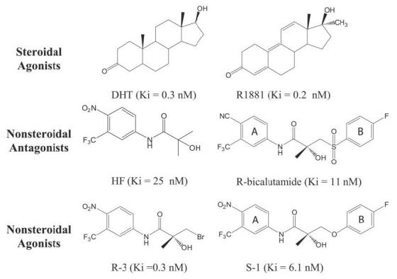
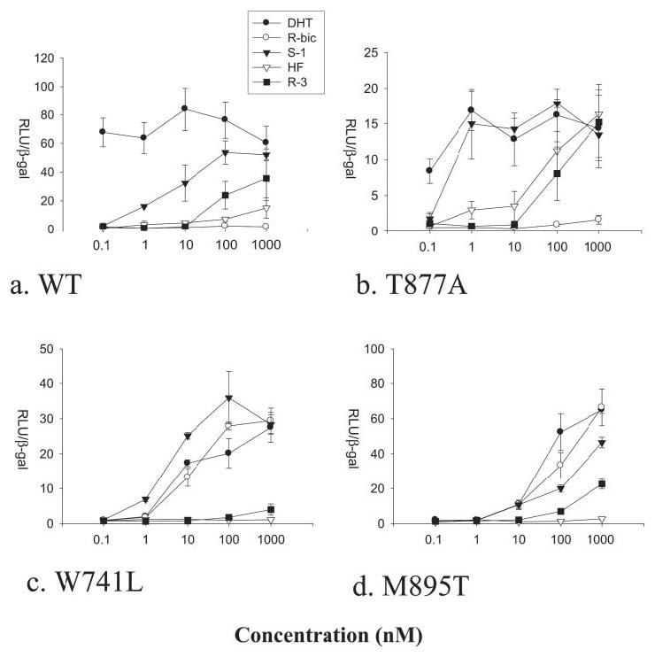
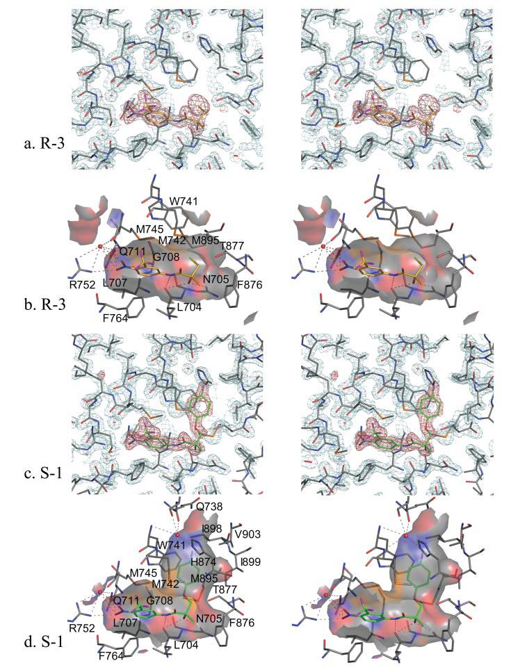
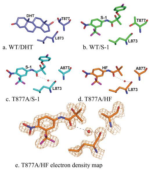
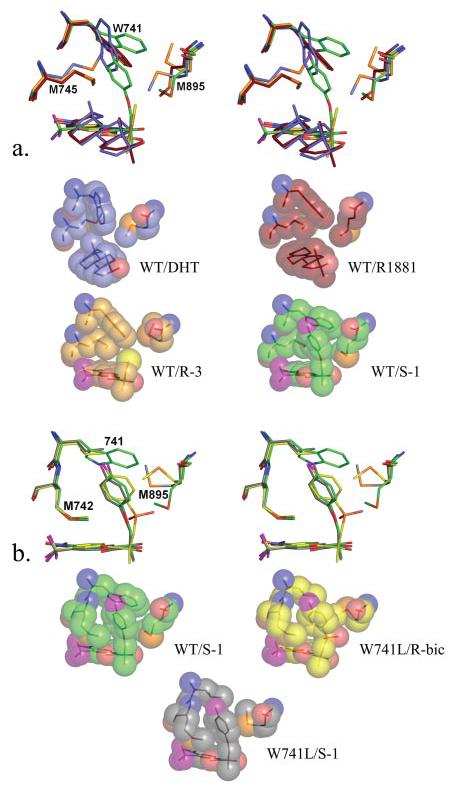
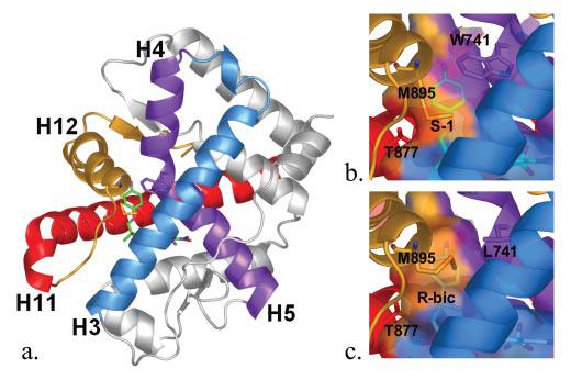
The mechanism by which the androgen receptor (AR) distinguishes between agonist and antagonist ligands is poorly understood. AR antagonists are currently used to treat prostate cancer. However, mutations commonly develop in patients that convert these compounds to agonists. Recently, our laboratory discovered selective androgen receptor modulators, which structurally resemble the nonsteroidal AR antagonists bicalutamide and hydroxyflutamide but act as agonists for the androgen receptor in a tissue-selective manner. To investigate why subtle structural changes to both the ligand and the receptor (i.e. mutations) result in drastic changes in activity, we studied structure-activity relationships for nonsteroidal AR ligands through crystallography and site-directed mutagenesis, comparing bound conformations of R-bicalutamide, hydroxyflutamide, and two previously reported nonsteroidal androgens, S-1 and R-3. These studies provide the first crystallographic evidence of the mechanism by which nonsteroidal ligands interact with the wild type AR. We have shown that changes induced to the positions of Trp-741, Thr-877, and Met-895 allow for ligand accommodation within the AR binding pocket and that a water-mediated hydrogen bond to the backbone oxygen of Leu-873 and the ketone of hydroxyflutamide is present when bound to the T877A AR variant. Additionally, we demonstrated that R-bicalutamide stimulates transcriptional activation in AR harboring the M895T point mutation. As a whole, these studies provide critical new insight for receptor-based drug design of nonsteroidal AR agonists and antagonists.
Figures
Similar articles
-
Molecular mechanism of R-bicalutamide switching from androgen receptor antagonist to agonist induced by amino acid mutations using molecular dynamics simulations and free energy calculation.J Comput Aided Mol Des. 2016 Dec;30(12):1189-1200. doi: 10.1007/s10822-016-9992-2. Epub 2016 Nov 15. J Comput Aided Mol Des. 2016. PMID: 27848066
-
Interaction mechanism exploration of R-bicalutamide/S-1 with WT/W741L AR using molecular dynamics simulations.Mol Biosyst. 2015 Dec;11(12):3347-54. doi: 10.1039/c5mb00499c. Epub 2015 Oct 7. Mol Biosyst. 2015. PMID: 26442831
-
The transcriptional co-activator cAMP response element-binding protein-binding protein is expressed in prostate cancer and enhances androgen- and anti-androgen-induced androgen receptor function.Am J Pathol. 2003 Jan;162(1):233-41. doi: 10.1016/S0002-9440(10)63814-X. Am J Pathol. 2003. PMID: 12507906 Free PMC article.
-
Structural features discriminate androgen receptor N/C terminal and coactivator interactions.Mol Cell Endocrinol. 2012 Jan 30;348(2):403-10. doi: 10.1016/j.mce.2011.03.026. Epub 2011 Jun 1. Mol Cell Endocrinol. 2012. PMID: 21664945 Free PMC article. Review.
-
Antiandrogens: selective androgen receptor modulators.Mol Cell Endocrinol. 2002 Dec 30;198(1-2):97-103. doi: 10.1016/s0303-7207(02)00373-8. Mol Cell Endocrinol. 2002. PMID: 12573819 Review.
Cited by
-
Bioinformatics and variability in drug response: a protein structural perspective.J R Soc Interface. 2012 Jul 7;9(72):1409-37. doi: 10.1098/rsif.2011.0843. Epub 2012 May 2. J R Soc Interface. 2012. PMID: 22552919 Free PMC article. Review.
-
Selective androgen receptor modulators in preclinical and clinical development.Nucl Recept Signal. 2008;6:e010. doi: 10.1621/nrs.06010. Epub 2008 Nov 26. Nucl Recept Signal. 2008. PMID: 19079612 Free PMC article. Review.
-
Apoptosis-mediated antiproliferation of A549 lung cancer cells mediated by Eugenia aquea leaf compound 2',4'-dihydroxy-6'-methoxy-3',5'-dimethylchalcone and its molecular interaction with caspase receptor in molecular docking simulation.Oncol Lett. 2020 May;19(5):3551-3557. doi: 10.3892/ol.2020.11466. Epub 2020 Mar 19. Oncol Lett. 2020. PMID: 32269629 Free PMC article.
-
Development of β-amino-carbonyl compounds as androgen receptor antagonists.Acta Pharmacol Sin. 2014 May;35(5):664-73. doi: 10.1038/aps.2013.201. Acta Pharmacol Sin. 2014. PMID: 24786235 Free PMC article.
-
Conformational dynamics of androgen receptors bound to agonists and antagonists.Sci Rep. 2021 Aug 5;11(1):15887. doi: 10.1038/s41598-021-94707-2. Sci Rep. 2021. PMID: 34354111 Free PMC article.
References
-
- Negro-Vilar A. J. Clin. Endocrinol. Metab. 1999;84:3459–3462. - PubMed
-
- Hara T, Miyazaki J, Araki H, Yamaoka M, Kanzaki N, Kusaka M, Miyamoto M. Cancer Res. 2003;63:149–153. - PubMed
-
- Veldscholte J, Berrevoets CA, Ris-Stalpers C, Kuiper GG, Jenster G, Trapman J, Brinkmann AO, Mulder E. J. Steroid Biochem. Mol. Biol. 1992;41:665–669. - PubMed
-
- Suzuki H, Akakura K, Komiya A, Aida S, Akimoto S, Shimazaki J. Prostate. 1996;29:153–158. - PubMed
Publication types
MeSH terms
Substances
Grants and funding
LinkOut - more resources
Full Text Sources
Other Literature Sources
Molecular Biology Databases
Research Materials
Miscellaneous
