. 2005 Sep 6;102(36):12684-9.
doi: 10.1073/pnas.0505975102. Epub 2005 Aug 29.

Three-dimensional structure of human monoamine oxidase A (MAO A): relation to the structures of rat MAO A and human MAO B

Affiliations

Three-dimensional structure of human monoamine oxidase A (MAO A): relation to the structures of rat MAO A and human MAO B

Luigi De Colibus et al. Proc Natl Acad Sci U S A. .

Abstract

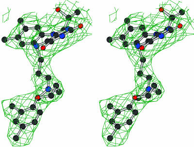
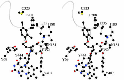
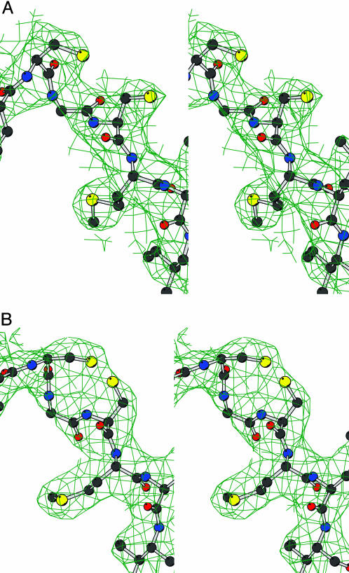
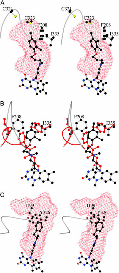
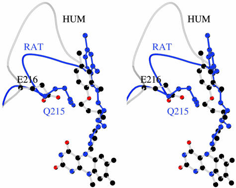
The three-dimensional structure of recombinant human monoamine oxidase A (hMAO A) as its clorgyline-inhibited adduct is described. Although the chain-fold of hMAO A is similar to that of rat MAO A and human MAO B (hMAO B), hMAO A is unique in that it crystallizes as a monomer and exhibits the solution hydrodynamic behavior of a monomeric form rather than the dimeric form of hMAO B and rat MAO A. hMAO A's active site consists of a single hydrophobic cavity of approximately 550 A3, which is smaller than that determined from the structure of deprenyl-inhibited hMAO B (approximately 700 A3) but larger than that of rat MAO A (approximately 450 A3). An important component of the active site structure of hMAO A is the loop conformation of residues 210-216, which differs from that of hMAO B and rat MAO A. The origin of this structural alteration is suggested to result from long-range interactions in the monomeric form of the enzyme. In addition to serving as a basis for the development of hMAO A specific inhibitors, these data support the proposal that hMAO A involves a change from the dimeric to the monomeric form through a Glu-151 --> Lys mutation that is specific of hMAO A [Andrès, A. M., Soldevila, M., Navarro, A., Kidd, K. K., Oliva, B. & Bertranpetit, J. (2004) Hum. Genet. 115, 377-386]. These considerations put into question the use of MAO A from nonhuman sources in drug development for use in humans.


Figures

Fig. 1.
Fig. 1.
Stereoview of the final 2FoFc electron density map for the clorgyline inhibitor covalently bound to the flavin in the X1 crystal form. The contour level is 1σ. The map was calculated with phases obtained by combining the phases obtained from inversion of the multiple-crystal 4-fold averaged electron density and the phases calculated from the refined model. Blue, nitrogens; black, carbons; red, oxygens; yellow, sulfurs.
Fig. 2.
Fig. 2.
Overall structure of hMAO A. The orientation is as in Fig. 1. (A) Ribbon representation of the monomer. The FAD-binding domain (residues 13–88, 220–294, and 400–462) is in blue; the substrate-binding domain (89–219 and 295–399) is in red; and the C-terminal membrane region (463–506) is in green. Residues 1–12, 111–115, and 507–527 are not visible in the electron density map. A dashed line connects residues 110–116. FAD and clorgyline are depicted in yellow and cyan ball-and-stick representation, respectively. The active site cavity-shaping loop 210–216 is depicted as black coil. (B) Stereoview of the superposition of the Cα traces of human MAO A (black) and MAO B (red). FAD and clorgyline bound to MAO A are shown as black ball-and-stick. Loop 210–216 of hMAO A and the equivalent loop 201–206 of hMAO B are shown as thick coils to highlight their different conformations.
Fig. 3.
Fig. 3.
Stereo closed-up view of the clorgyline site in hMAO A. Atom colors are as in Fig. 1. The backbone trace of loop 210–216 is shown as a coil.
Fig. 4.
Fig. 4.
Stereoview of the final 2FoFc electron density map for the Cys-321–Cys-323 pair in the X2 (A) and X1 (B) crystal forms. The maps were calculated as in Fig. 1, and the level was contoured at 1σ. Atom colors are as in Fig. 1.
Fig. 5.
Fig. 5.
Active site cavities in hMAO A and hMAO B. (A) The surface of active site cavity in hMAO A is shown in red chicken-wire representation in the same orientation as in Fig. 3. Clorgyline is depicted in black. (B) Active site comparison of hMAO A and hMAO B with the crucial Phe-208 and Ile-335 residues of hMAO A superimposed to the corresponding Ile-199 and Tyr-326 residues of hMAO B. The protein and inhibitor atoms of hMAO B are in red. With respect to A, the model has been rotated by ≈90° around the vertical axis in the plane of the drawing. (C) The active site cavity (red surface) of hMAO B in complex with deprenyl (black) is depicted in the same orientation as in A.
Fig. 6.
Fig. 6.
The different clorgyline binding modes in hMAO A and rMAO A. Generated by superposing the Cα atoms of the two MAO A structures, the stereoview highlights the different conformations of the bound inhibitor and of residues of the active site cavity-shaping loop 210–216. Atoms of rMAO A are in blue. The orientation is the same as in Fig. 3.

References

    1. Brunner, H. G., Nelen, M. R., Zandvoort, P., Abeling, N., Gennip, A. H., Wolter, E. C., Kuiper, M. A., Roper, H. H. & Oust, B. A. (1993) Am. J. Hum. Genet. 52, 578–580. - PMC - PubMed
    1. Caspi, A., McClay, J., Moffitt, T. E., Mill, J., Martin, J., Craig, I. W., Taylor, A. & Poulton, R. (2002) Science 297, 851–853. - PubMed
    1. Binda, C., Newton-Vinson, P., Hubálek, F., Edmondson, D. E. & Mattevi, A. (2002) Nat. Struct. Biol. 9, 22–26. - PubMed
    1. Binda, C., Li, M., Hubálek, F., Restelli, N., Edmondson, D. E. & Mattevi, A. (2003) Proc. Natl. Acad. Sci. USA 100, 9750–9755. - PMC - PubMed
    1. Ma, J., Yoshimura, M., Yamashita, E., Nakagawa, A., Ito, A. & Tsukihara, T. (2004) J. Mol. Biol. 338, 103–114. - PubMed

Publication types

Substances

LinkOut - more resources