. 2005 Sep 13;102(37):13254-9.
doi: 10.1073/pnas.0504830102. Epub 2005 Aug 29.

Microbial regulation of intestinal radiosensitivity

Affiliations

Microbial regulation of intestinal radiosensitivity

Peter A Crawford et al. Proc Natl Acad Sci U S A. .

Abstract

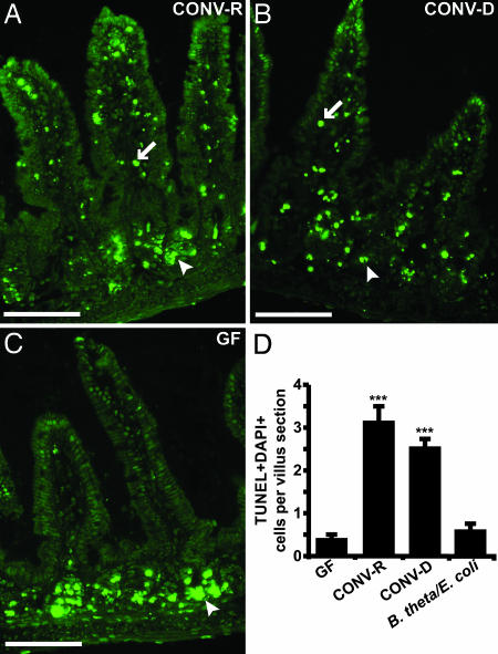
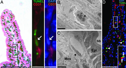
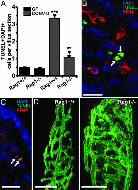
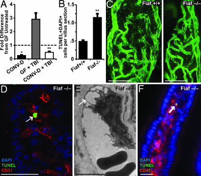
We describe a method for treating germ-free (GF) mice with gamma-irradiation and transplanting them with normal or genetically manipulated bone marrow while maintaining their GF status. This approach revealed that GF mice are markedly resistant to lethal radiation enteritis. Furthermore, administering lethal doses of total body irradiation to GF mice produces markedly fewer apoptotic endothelial cells and lymphocytes in the mesenchymal cores of their small intestinal villi, compared with conventionally raised animals that have acquired a microbiota from birth. Analysis of GF and conventionally raised Rag1-/- mice disclosed that mature lymphocytes are not required for the development of lethal radiation enteritis or the microbiota-associated enhancement of endothelial radiosensitivity. Studies of gnotobiotic knockout mice that lack fasting-induced adipose factor (Fiaf), a fibrinogen/angiopoietin-like protein normally secreted from the small intestinal villus epithelium and suppressed by the microbiota, showed that Fiaf deficiency results in loss of resistance of villus endothelial and lymphocyte populations to radiation-induced apoptosis. Together, these findings provide insights about the cellular and molecular targets involved in microbial regulation of intestinal radiosensitivity.


Figures

Fig. 1.
Fig. 1.
Microbiota-associated sensitivity of villus core mesenchymal cells to TBI-induced apoptosis. TUNEL+ cells appearing 4 h after 16 Gy of TBI in the distal small intestines of CONV-R (A), CONV-D (B), and GF (C)WTFVB/N mice. Fluorescence microscopy reveals green TUNEL+ cells in the crypt epithelium (arrowheads) of all three mice and in the villus core mesenchyme (arrows) of CONV-R and CONV-D mice. In contrast, TUNEL+ cells are absent from the villus core mesenchyme of the GF animal. (Scale bars, 50 μm.) (D) The mean number (±1 SD) of TUNEL+/DAPI+ mesenchymal cells per distal small intestinal villus sections prepared from GF, CONV-R, CONV-D, and B. theta/E. coli-biassociated FVB/N mice (four to five mice scored per group; ≥100 villus sections assayed per animal). ***, P < 0.001, compared with GF. In nonirradiated CONV-R controls, the mean number of apoptotic cells per section of villus core mesenchyme was ≤0.2.
Fig. 2.
Fig. 2.
Apoptosis of CD31+ endothelial cells and CD45+ leukocytes in the villus mesenchyme of a CONV-R mouse treated with 16 Gy of TBI. (A) Small intestinal villus section stained with hematoxylin and eosin (Left). (Scale bar, 100 μm.) The expanded views of the boxed region (Right) show an adjacent section after staining with TUNEL reagents and antibodies to CD31. The arrow points to a TUNEL+ nucleus in a CD31+ endothelial cell. (Scale bar, 10 μm.) (B) TEM image of a mesenchymal endothelial cell (arrowhead) from the distal small intestine of a CONV-R mouse 4 h after 16 Gy of TBI. Nuclear condensation and cellular protrusion into the capillary lumen are characteristics of endothelial apoptosis. (Scale bar, 2 μm.) (C) A TEM image of a representative villus mesenchymal endothelial cell (arrowhead) from the distal small intestine of a GF B6 mouse killed 4 h after 16 Gy TBI. The cell has normal morphology. Epi, epithelium; Mes, mesenchyme, bb, brush border on the apical surface of enterocytes. (Scale bar, 4 μm.) (D) A single image from a 3D reconstruction of serial 1-μm-thick scans of a villus section from a CONV-R mouse killed 4 h after 16 Gy of TBI. Arrows denote TUNEL+ CD45+ cells, whereas arrowheads denote TUNEL+ CD45- cells. CD45 is a surface marker, whereas TUNEL stains nuclei: therefore, TUNEL+ cells that coexpress CD45 will not appear yellow. (Scale bar, 80 μm.) (Inset) A higher-power view of the boxed area.
Fig. 3.
Fig. 3.
Rag1-/- mice reveal that the microbiota-dependent apoptotic response of villus endothelial cells to 16 Gy of TBI does not require mature T or B cells. (A) Quantitative assessment of TUNEL+ DAPI+ cells in the villus mesenchyme of B6 GF and CONV-D WT and Rag1-/- mice killed 4 h after 16 Gy of TBI (n = 4–6 mice per group; ≥100 villus sections assayed per mouse). ***, P < 0.01, compared with GF Rag1+/+ mice; **, P < 0.01, compared with GF Rag1-/- mice; Ψ, P < 0.05, compared with CONV-D Rag1+/+ mice. (B) Two TUNEL+ CD31+ endothelial cells (arrows) in the small intestinal villus mesenchyme of a CONV-D Rag1-/- mouse treated as in A. The image was acquired from a 0.5-μm-thick digital scan of the section. (C) Adjacent section showing two TUNEL+ CD45+ leukocytes (arrows) in the villus mesenchyme. (D) Microscopic angiograms showing single images from 3D reconstructions of serial 1-μm-thick scans of nonirradiated distal small intestinal villi from CONV-D Rag1+/+ and Rag1-/- distal small intestinal villi. The complexity of the submucosal capillary network present in the villus core mesenchyme can be assessed by noting the size of open areas (“windows”) circumscribed by interconnected vessels. Analysis of this parameter indicated that network complexity was not appreciably different between Rag1-/- and WT mice with the two genotypes. (Scale bars, 20 μm.)
Fig. 4.
Fig. 4.
Loss of Fiaf results in loss of resistance to TBI-induced apoptosis in GF mice. (A) Quantitative RT-PCR analysis shows that conventionalization of GF WT mice suppresses Fiaf expression in the distal small intestine and that this effect is not abrogated after 16 Gy of TBI (mean values ± 1 SD are plotted; n = 4 mice per group; *, P < 0.05, compared with GF untreated B6 mice; **, P < 0.01, compared with GF mice treated with 16 Gy of TBI 4 h before they were killed). (B) Quantitative assessment of apoptosis in the villus mesenchyme of GF Fiaf-/- mice and their WT littermates 4 h after 18 Gy of TBI (n = 9 mice per group; ≥100 villus sections assayed per mouse; mean ± 1 SD; **, P < 0.01, compared with GF Fiaf+/+). (C) Single images from digital 3D reconstructions of small intestinal villus capillary networks in GF Fiaf-/- and their WT littermates. (Scale bars, 20 μm.) (D) TUNEL+ CD31+ endothelial cell (arrow) in the small intestinal villus mesenchyme of a GF Fiaf-/- mouse treated as in A. (Scale bar, 50 μm.) (E) TEM image of a villus mesenchymal endothelial cell in a Fiaf-/- mouse killed 4 h after 18 Gy of TBI. Note the nuclear protrusion and avulsion from the basement membrane (arrow), two ultrastructural manifestations of endothelial cell death. (Scale bar, 5 μm.) (F) TUNEL+ CD45+ leukocyte (arrow) in the small intestinal villus mesenchyme of a GF Fiaf-/- mouse treated as in A. (Scale bar, 20 μm.)

References

    1. Eckburg, P. B., Bik, E. M., Bernstein, C. N., Purdom, E., Dethlefsen, L., Sargent, M., Gill, S. R., Nelson, K. E. & Relman, D. A. (2005) Science 308, 1635-1638. - PMC - PubMed
    1. Bäckhed, F., Ley, R. E., Sonnenburg, J. L., Peterson, D. A. & Gordon, J. I. (2005) Science 307, 1915-1920. - PubMed
    1. Bäckhed, F., Ding, H., Wang, T., Hooper, L. V., Koh, G. Y., Nagy, A., Semenkovich, C. F. & Gordon, J. I. (2004) Proc. Natl. Acad. Sci. USA 101, 15718-15723. - PMC - PubMed
    1. Savage, D. C. (1986) Annu. Rev. Nutr. 6, 155-178. - PubMed
    1. Hooper, L. V., Midtvedt, T. & Gordon, J. I. (2002) Annu. Rev. Nutr. 22, 283-307. - PubMed

Publication types

MeSH terms