. 2005 Sep 6;102(36):12736-41.
doi: 10.1073/pnas.0501808102. Epub 2005 Aug 29.

Structure of AMA1 from Plasmodium falciparum reveals a clustering of polymorphisms that surround a conserved hydrophobic pocket

Affiliations

Structure of AMA1 from Plasmodium falciparum reveals a clustering of polymorphisms that surround a conserved hydrophobic pocket

Tao Bai et al. Proc Natl Acad Sci U S A. .

Abstract

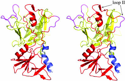
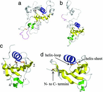
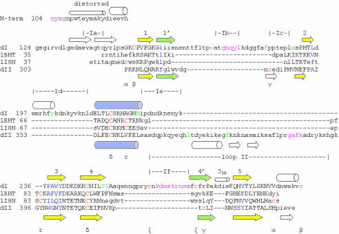
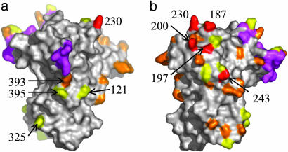
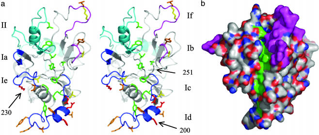
Apical membrane antigen 1 (AMA1) is a leading malaria vaccine candidate that possesses polymorphisms that may pose a problem for a vaccine based on this antigen. Knowledge of the distribution of the polymorphic sites on the surface of AMA1 is necessary to obtain a detailed understanding of their significance for vaccine development. For this reason we have sought to determine the three-dimensional structure of AMA1 using x-ray crystallography. The central two-thirds of AMA1 is relatively conserved among Plasmodium species as well as more distantly related apicomplexan parasites, and contains two clusters of disulfide-bonded cysteines termed domains I and II. The crystal structure of this fragment of AMA1 reported here reveals that domains I+II consists of two intimately associated PAN domains. PAN domain I contains many long loops that extend from the domain core and form a scaffold for numerous polymorphic residues. This extreme adaptation of a PAN domain reveals how malaria parasites have introduced significant flexibility and variation into AMA1 to evade protective human antibody responses. The polymorphisms on the AMA1 surface are exclusively located on one side of the molecule, presumably because this region of AMA1 is most accessible to antibodies reacting with the parasite surface. Moreover, the most highly polymorphic residues surround a conserved hydrophobic trough that is ringed by domain I and domain II loops. Precedents set by viral receptor proteins would suggest that this is likely to be the AMA1 receptor binding pocket.


Figures

Fig. 1.
Fig. 1.
Stereo view of the AMA1 domain I+II structure showing the two interconnected domains. The 20 aa from the N-terminal extension are colored blue, domain I is yellow, domain II is red, and loops that are disordered in the structure are violet. This and all other figures depicting the structure were generated by using pymol (www.pymol.org).
Fig. 2.
Fig. 2.
The PAN domains of AMA1 domain I (a) and AMA1 domain II (b), N domain of hepatocyte growth factor (c) [1BHT (47)], and the apple domain of leech antiplatelet protein (d) [1I8N (48)]. Disulfide linkages are shown in orange. The numbering of the secondary structure elements is shown in c with the corresponding numbering in the primary sequence in Fig. 3 according to the scheme of Ultsch et al. (47). Characteristic PAN or apple domain disulfides are shown in d. Disordered loops in the AMA1 structure are colored violet.
Fig. 3.
Fig. 3.
Sequence alignment based on the tertiary alignment of PAN domain crystal structures. Carbon alphas that align within 2.5 Å in the tertiary structures are shown in uppercase. N-term, the N-terminal 20 aa; dI, domain I of AMA1; dII, domain II of AMA1; 1BHT, N domain of hepatocyte growth factor (47); 1I8N, leach anti-platelet protein (48). Central PAN domain sheets are colored yellow, the PAN helix is blue, and the peripheral sheets are green, with secondary structure elements numbered as in Fig. 2. Residues in violet are disordered in the structure. Cysteines are colored orange. The same Greek letter below two cysteines indicates that they are disulfide-bonded. Residues at PAN-domain conserved hydrophobic positions are colored blue. Surface-exposed hydrophobic Plasmodium-conserved residues that line the base of the hydrophobic trough are colored green. The N-terminal “distorted” helix is helical but contains carbonyls that interact with waters, side chains, and main-chain nitrogens in 310 and α-helical connections. The short 310 helix is shown as a small cylinder. α-helices are shown as large cylinders, and β-strands as arrows. Positions of domain I loops, Ia to If, are indicated together with the domain II loop (loop II).
Fig. 4.
Fig. 4.
Clustering of polymorphic residues on the surface of AMA1. Side chains 197, 200, 230, and 243, shown in red, are highly polymorphic. High-frequency dimorphisms are shown in orange, with low-frequency dimorphisms in yellow, as described in Materials and Methods. (a) Surface view of the “nonpolymorphic face.” (b) Surface view of the “polymorphic face.” Areas colored violet are disordered in the structure.
Fig. 5.
Fig. 5.
AMA1 loops surround a conserved hydrophobic trough. (a) Stereo view with surface-exposed Plasmodium-conserved hydrophobic residues that line the base of the trough shown in green. Tyrosine 251, located in the trough center, is identical in all apicomplexan sequences. Polymorphic sites are shown in red, with K230 and H200 indicated. Dimorphic sites are colored orange (high frequency) and yellow (low frequency). “Ordered” loops are colored blue with the domain I loops Ia, Ic, Id, and Ie in dark blue and the domain II loop in light blue. Disordered loops Ib and If are colored violet. The disordered loops contain four dimorphic sites, and the approximate location of these residues is indicated. (b) Surface view of the hydrophobic trough. Carbon and sulfur atoms that form the base of the hydrophobic trough are colored green. Other carbon and sulfur atoms are colored white. Oxygen atoms are colored red, and nitrogen atoms are blue. Disordered regions are colored violet.

References

    1. Breman, J. G., Alilio, M. S. & Mills, A. (2004) Am. J. Trop. Med. Hyg. 71, 1–15. - PubMed
    1. Sachs, J. & Malaney, P. (2002) Nature 415, 680–685. - PubMed
    1. Baird, J. K. (1995) Parasitol. Today 11, 105–111. - PubMed
    1. Cohen, S., McGregor, I. A. & Carrington, S. C. (1961) Nature 192, 733–737. - PubMed
    1. Cohen, S. & Butcher, G. A. (1970) Nature 225, 732–734. - PubMed

Publication types

MeSH terms

Associated data