. 2005 Sep 6;102(36):12724-9.
doi: 10.1073/pnas.0506124102. Epub 2005 Aug 29.

Specificity versus stability in computational protein design

Affiliations

Specificity versus stability in computational protein design

Daniel N Bolon et al. Proc Natl Acad Sci U S A. .

Abstract

Protein-protein interactions can be designed computationally by using positive strategies that maximize the stability of the desired structure and/or by negative strategies that seek to destabilize competing states. Here, we compare the efficacy of these methods in reengineering a protein homodimer into a heterodimer. The stability-design protein (positive design only) was experimentally more stable than the specificity-design heterodimer (positive and negative design). By contrast, only the specificity-design protein assembled as a homogenous heterodimer in solution, whereas the stability-design protein formed a mixture of homodimer and heterodimer species. The experimental stabilities of the engineered proteins correlated roughly with their calculated stabilities, and the crystal structure of the specificity-design heterodimer showed most of the predicted side-chain packing interactions and a main-chain conformation indistinguishable from the wild-type structure. These results indicate that the design simulations capture important features of both stability and structure and demonstrate that negative design can be critical for attaining specificity when competing states are close in structure space.


Figures

Fig. 1.
Fig. 1.
SspB dimer interface. (A) Ribbon diagram of SspB from the wild-type crystal structure (16) with one subunit colored purple and the other subunit colored blue. (BD) Molecular images of the SspB dimer interface showing a transparent surface of strand β7 and helix α1. Side chains that were allowed to vary in design calculations are shown in space-fill representation. Nearby side chains whose geometries did not vary during the calculations are shown in stick representation. (B) Wild-type LAYV/LAYV interface. (C) Stability-design FAFI/LALI interface. (D) Specificity-design LSLA/YGFM interface.
Fig. 2.
Fig. 2.
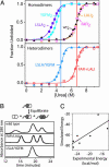
Heterodimer and homodimer stabilities. (A) Urea-induced unfolding of SspB dimers was monitored by changes in tryptophan fluorescence at 30°C in 50 mM potassium phosphate (pH 6.8). (Upper) Unfolding transitions for the LSLA, YGFM, wild-type, FAFI, and LALI homodimers. (Lower) Unfolding transitions for the LSLA/YGFM and FAFI+LALI proteins. For comparison between homodimers and heterodimers, vertical blue and red lines mark the Cm for LSLA/YGFM and FAFI+LALI, respectively. Fitted ΔG and m values are listed in Table 2. Based on these values, an equimolar mixture of LSLA and YGFM subunits results in 99% of the LSLA/YGFM heterodimer at equilibrium. Denaturation experiments were fit to a two-state model in which native dimers are in equilibrium with unfolded monomers (25). This model predicts that denaturation should be concentration-dependent and that denaturation monitored by circular dichroism should give the same transition. Both predictions were experimentally confirmed for the LSLA homodimer, the protein of lowest stability (data not shown). (B) Exchange reactions. Heterodimers containing one full-length subunit and one truncated subunit were purified by ion-exchange chromatography, incubated for 24 h, and then rechromatographed. The wild-type heterodimer and FAFI/LALI heterodimer equilibrated to form a mixture of both homodimers and the heterodimer. The LSLA/YGFM heterodimer did not form appreciable quantities of either homodimer. (C) Energies calculated from the design simulations (Table 1) are plotted against the experimental ΔG values determined by urea denaturation (Table 2). The simulated energies were calculated for interactions of the optimized positions in the folded state and were intended to capture the relative stability of the sequence variants. However, these calculations do not include terms for main-chain to main-chain interactions or folding entropy and therefore are not intended to represent absolute stability. The line is a linear fit (R = 0.78).
Fig. 3.
Fig. 3.
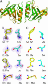
Crystal structure of LSLA/YGFM heterodimer. (A) Ribbon representation of an alignment of the LSLA/YGFM heterodimer structure (Protein Data Bank entry 1ZSZ; green) with the wild-type SspB homodimer structure (Protein Data Bank entry 1OU9; orange). The main-chain RMSD between these structures is 0.4 Å, indicating that the redesigned dimer interface does not perturb the overall structure. (B)2FcFo simulated annealing omit maps of electron density for designed mutations at the dimer interface contoured to 1.0 σ. (C) Side-chain geometries of optimized amino acids in the designed heterodimer model are generally similar to those observed in the crystal structure.

References

    1. Dahiyat, B. I. & Mayo, S. L. (1997) Science 278, 82–87. - PubMed
    1. Bolon, D. N. & Mayo, S. L. (2001) Proc. Natl. Acad. Sci. USA 98, 14274–14279. - PMC - PubMed
    1. Looger, L. L., Dwyer, M. A., Smith, J. J. & Hellinga, H. W. (2003) Nature 423, 185–190. - PubMed
    1. Kuhlman, B., Dantas, G., Ireton, G. C., Varani, G., Stoddard, B. L. & Baker, D. (2003) Science 302, 1364–1368. - PubMed
    1. Wilson, C., Mace, J. E. & Agard, D. A. (1991) J. Mol. Biol. 220, 495–506. - PubMed

Publication types

MeSH terms

Substances

Associated data