. 2006 Jan 1;393(Pt 1):69-77.
doi: 10.1042/BJ20050959.

MAPK interacts with occludin and mediates EGF-induced prevention of tight junction disruption by hydrogen peroxide

Affiliations

MAPK interacts with occludin and mediates EGF-induced prevention of tight junction disruption by hydrogen peroxide

Shyamali Basuroy et al. Biochem J. .

Abstract

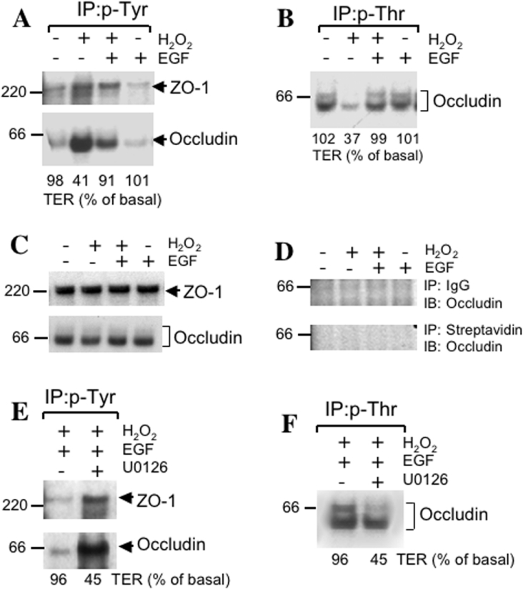
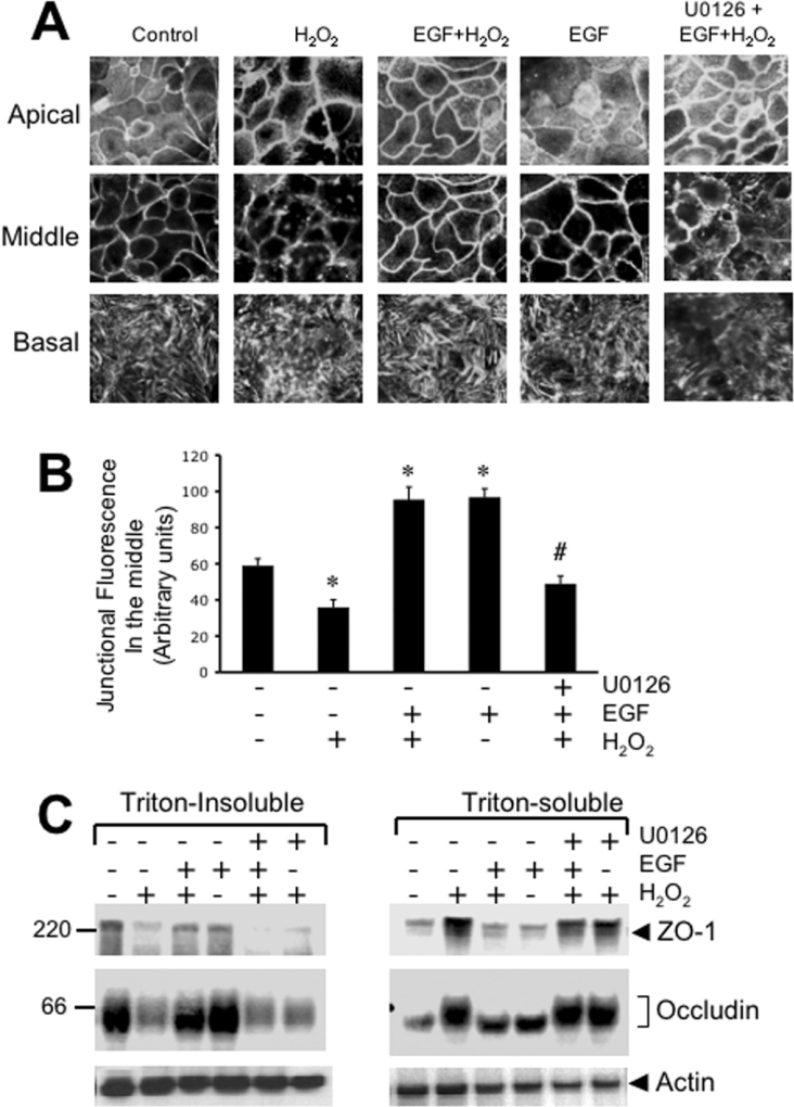
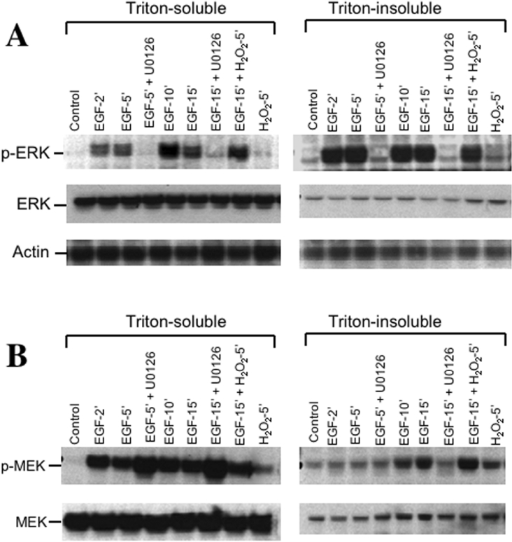
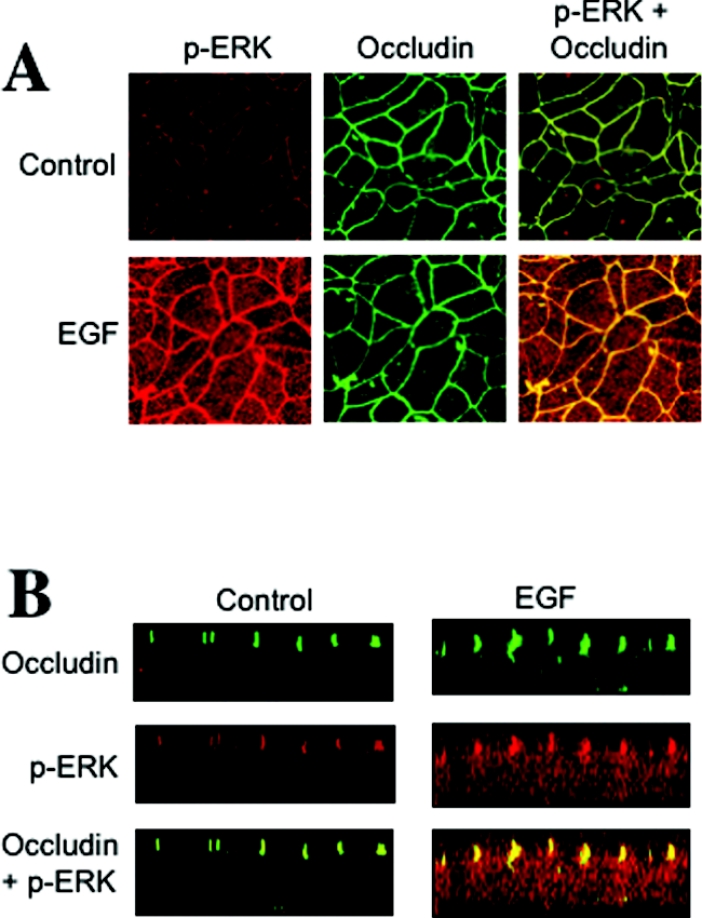
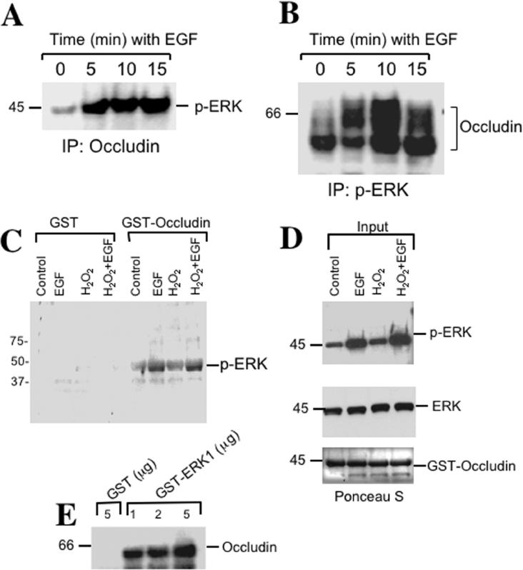
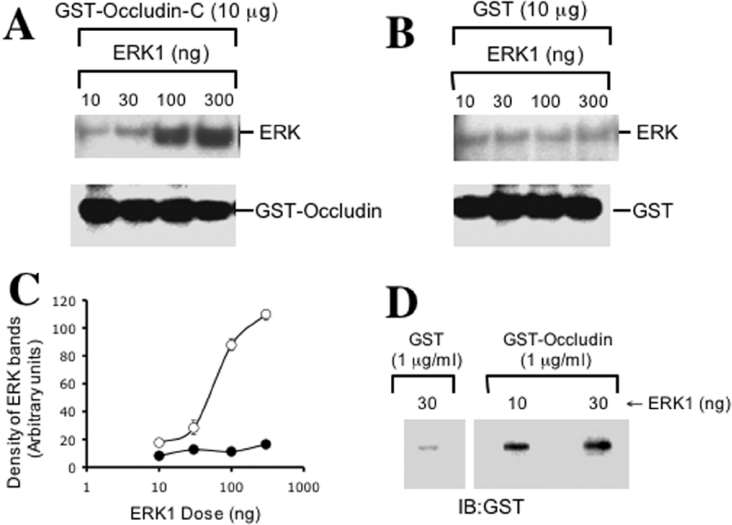
The MAPK (mitogen-activated protein kinase) pathway is a major intracellular signalling pathway involved in EGF (epithelial growth factor) receptor-mediated cell growth and differentiation. A novel function of MAPK activity in the mechanism of EGF-mediated protection of TJs (tight junctions) from H2O2 was examined in Caco-2 cell monolayers. EGF-mediated prevention of H2O2-induced increase in paracellular permeability was associated with the prevention of H2O2-induced Tyr-phosphorylation, Thr-dephosphorylation and cellular redistribution of occludin and ZO-1 (zonula occludin-1). EGF also prevented H2O2-induced disruption of the actin cytoskeleton and the dissociation of occludin and ZO-1 from the actin-rich detergent-insoluble fractions. MEK (MAPK/ERK kinase, where ERK stands for extracellular signal related kinase) inhibitors, PD98059 and U0126, completely blocked these protective effects of EGF on TJs. EGF rapidly increased the levels of phosphorylated MEK (p-MEK) in detergent-soluble fractions and phosphorylated ERK (p-ERK) in detergent-insoluble fractions. p-ERK was colocalized and co-immunoprecipitated with occludin. GST (glutathione S-transferase) pull-down assay showed that the C-terminal tail of occludin binds to p-ERK in Caco-2 cell extracts. Pair-wise binding studies using recombinant proteins demonstrated that ERK1 directly interacts with the C-terminal tail of occludin. Therefore the present study shows that ERK interacts with the C-terminal region of occludin and mediates the prevention of H2O2-induced disruption of TJs by EGF.


Figures

Figure 1
Figure 1. EGF prevents H2O2-induced increases in paracellular permeability and redistribution of occludin and ZO-1 by a MAPK-dependent mechanism
Caco-2 cell monolayers were incubated with or without 20 μM H2O2 in the presence or absence of 30 nM EGF for 3 h. In some groups cell monolayers were pretreated with PD98059 (30 μM) or U0126 (10 μM) for 1 h before administration of EGF and H2O2. TER (A) was recorded at various times and values at 3 h were calculated as percentage of basal values. Unidirectional flux of FITC-inulin (B) measured during 3 h of treatment with H2O2. Values are mean±S.E.M. (n=8). *, indicates the values that are significantly different (P<0.05) from values for H2O2; #, indicates the values that are significantly different from values for the EGF+H2O2 group. (C) Caco-2 cell monolayers were incubated with H2O2 in the presence or absence of EGF and in the presence or absence of U0126. After 3 h treatment, cell monolayers were fixed and double-labelled for occludin and ZO-1 by immunofluorescence methods. Fluorescence images were collected by confocal microscopy.
Figure 2
Figure 2. EGF prevents H2O2-induced alteration in phosphorylation of occludin and ZO-1 by a MAPK-dependent mechanism
(A) and (B) Caco-2 cell monolayers were incubated with or without H2O2 in the presence or absence of 30 nM EGF for 3 h. Proteins extracted under denatured conditions were subjected to immunoprecipitation of p-Tyr (A) or p-Thr (B) followed by immunoblot analysis for occludin and ZO-1. Values at the bottom of immunoblots represent the TER values for the corresponding experiment. This experiment was repeated twice with similar results. (C) and (D) Caco-2 cell monolayers were incubated as described above for (A) and (B). Tissue extracts were either directly immunoblotted for total occludin and ZO-1 (C) or control-immunoprecipitation was performed using preimmune rabbit-IgG with Protein A–Sepharose or streptavidin–agarose followed by immunoblot analysis for occludin and ZO-1. (E) and (F) Caco-2 cell monolayers were incubated with EGF and H2O2 in the presence or absence of U0126 for 3 h. Extracted proteins were subjected to immunoprecipitation of p-Tyr (E) or p-Thr (F) followed by immunoblot analysis for occludin and ZO-1. This experiment was repeated twice with similar results.
Figure 3
Figure 3. EGF prevents H2O2-induced reorganization of actin cytoskeleton and its interaction with TJ proteins by a MAPK-dependent mechanism
Caco-2 cell monolayers were incubated with or without H2O2 in the presence or absence of EGF and U0126 for 3 h. (A) Cell monolayers were fixed in paraformaldehyde and stained with Alexa Fluor™ 488-conjugated phalloidin. Fluorescence images from different depths in epithelium (apical, middle and basal) were obtained by confocal microscopy. (B) Semi-quantitative analysis of junctional fluorescence in the middle part of the cells. Intensity of fluorescence at the junctions was measured over a constant area of 1 mm2 and the values are presented as arbitrary units. Values are mean±S.E.M. (n=4; each value is the average of 4 different regions from the same image). *, indicate the values that are significantly different (P<0.05) from values for control cells; # indicates the values that are significantly different from values for the EGF+H2O2 group. (C) Triton-insoluble and Triton-soluble fractions were prepared from different cell monolayers and immunoblotted for occludin, ZO-1 and actin.
Figure 4
Figure 4. EGF activates ERK in Caco-2 cell monolayers by a MEK-dependent mechanism
Caco-2 cell monolayers were pretreated with or without U0126 for 1 h followed by incubation with EGF for various times. In additional cell monolayers, H2O2 was added 10 min after EGF and the incubation was continued for a further 5 min. Triton-insoluble and Triton-soluble fractions were prepared and immunoblotted for ERK and p-ERK (A), or MEK and p-MEK (B). Samples were also immunoblotted for actin as a house keeping protein (A).
Figure 5
Figure 5. p-ERK is co-localized with occludin at the TJ in Caco-2 cell monolayers
(A) Caco-2 cell monolayers were incubated in the presence or absence of EGF for 5 min. Cell monolayers were fixed in acetone/methanol and stained for occludin and p-ERK by immunofluorescence methods. (B) Optical z-sections of fluorescence images were collected by confocal microscopy.
Figure 6
Figure 6. Phospho-ERK interacts with TJ protein complex
(A) Caco-2 cell monolayers were incubated with EGF for various durations. Occludin was immunoprecipitated from proteins extracted from Triton-insoluble fractions under native conditions. Immunocomplexes were then immunoblotted for p-ERK. (B) After EGF treatment, p-ERK was immunoprecipitated from native protein extracts of the Triton-insoluble fractions and immunoblotted for occludin. (C) The C-terminal tail of occludin (150 amino acids) was expressed in E. coli as a GST fusion protein and incubated with Triton-soluble protein extracts from Caco-2 cells, with or without, EGF for 5 min and H2O2 for 30 min. GST–occludin was pulled-down with GSH–agarose and immunoblotted for p-ERK. Non-specific binding was determined by incubation of Triton-soluble protein extracts with GST. (D) Immunoblot analysis of p-ERK and ERK protein extracts used in (C). The blot was stained for total protein by Ponceau S dye to visualize the amount of GST–occludin. (E) GST, or various amounts of recombinant GST–ERK1 incubated with Triton-soluble protein extracts from untreated Caco-2 cells, and GSH–agarose pull-down were immunoblotted for occludin.
Figure 7
Figure 7. ERK1 directly interacts with the C-terminal region of occludin
(A) Various amounts of thrombin-cleaved recombinant ERK1 were incubated with GST–occludin-C. GSH–agarose pull-down was immunoblotted for ERK. Blot was also immunoblotted for GST to visualize the amount of GST–occludin used in the assay. (B) Binding assay was conducted as described in (A) using GST instead of GST–occludin to determine the non-specific binding of ERK1. (C) Densitometric analysis of ERK1 bands in panels A and B. Density is presented as arbitrary units. Values are mean±S.E.M. (n=3). (D) Thrombin-cleaved ERK1 was slot blotted on PVDF membrane and overlayed with GST–occludin-C or GST. Washed blots were then probed for GST.

References

    1. Trier J. S. Celiac sprue and refractory sprue. In: Sleisenger M., Fordstran J., editors. Gastrointestinal Disease. Philadelphia: W. B. Saunders; 1978. pp. 1029–1051.
    1. Hollander D. Permeability in Crohn's disease: altered barrier functions in healthy relatives? Gastroenterology. 1993;104:1848–1851. - PubMed
    1. Mathay M. A., Fukuda N., Frank J., Kallet R., Daniel B., Sakuma T. Alveolar epithelial barrier. Role in lung fluid balance in clinical lung injury. Clin. Chest Med. 2000;21:477–490. - PubMed
    1. Anderson J. M., van Italie C. M. Tight junctions and the molecular basis for regulation of paracellular permeability. Am. J. Physiol. 1995;269:G467–G475. - PubMed
    1. Tsukita S., Furuse M. Occludin and claudins in tight-junction strands: leading or supporting players? Trends Cell Biol. 1999;9:268–273. - PubMed

Publication types