. 2005 Nov;16(11):5202-14.
doi: 10.1091/mbc.e05-03-0256. Epub 2005 Aug 31.

Taz1, an outer mitochondrial membrane protein, affects stability and assembly of inner membrane protein complexes: implications for Barth Syndrome

Affiliations

Taz1, an outer mitochondrial membrane protein, affects stability and assembly of inner membrane protein complexes: implications for Barth Syndrome

Katrin Brandner et al. Mol Biol Cell. 2005 Nov.

Abstract

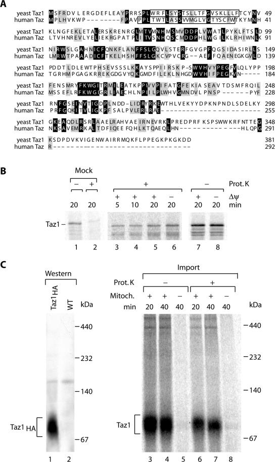
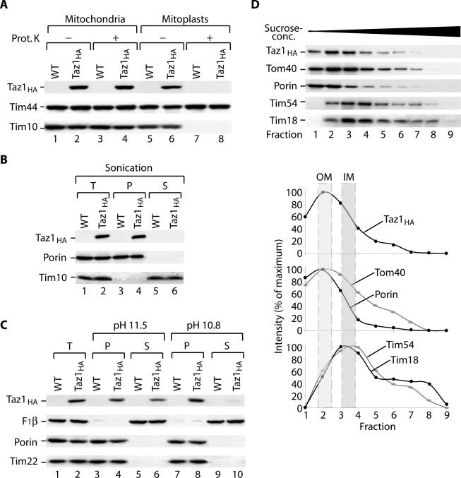
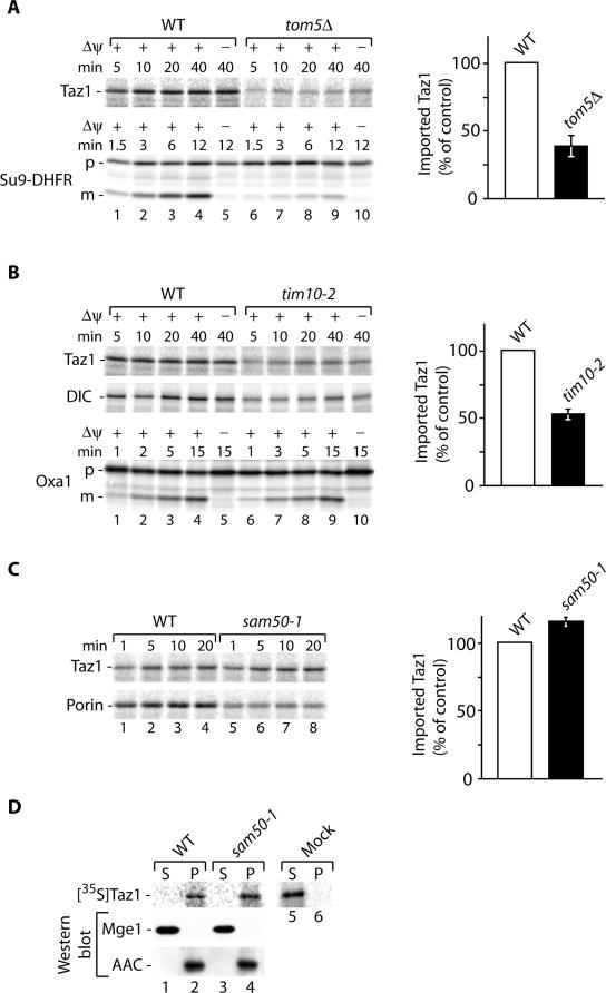
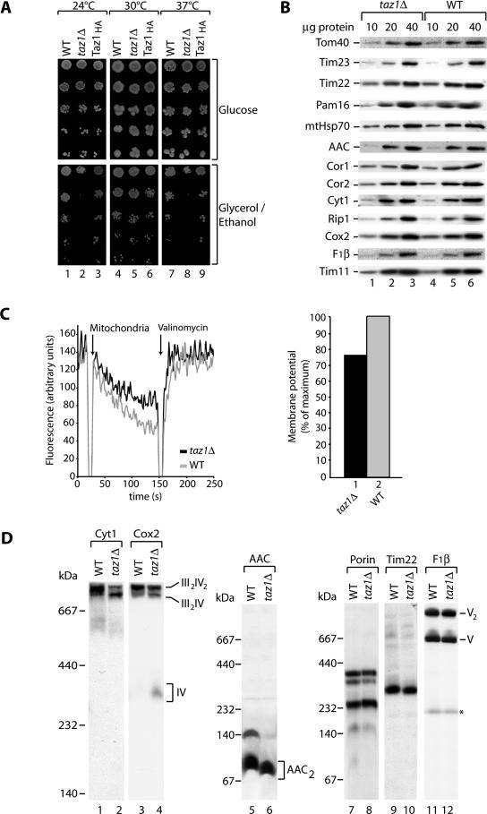
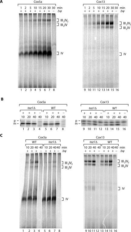
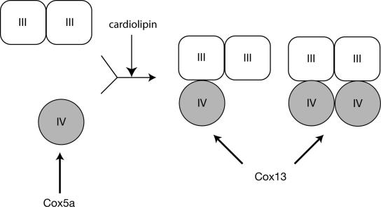
The Saccharomyces cerevisiae Taz1 protein is the orthologue of human Tafazzin, a protein that when inactive causes Barth Syndrome (BTHS), a severe inherited X-linked disease. Taz1 is a mitochondrial acyltransferase involved in the remodeling of cardiolipin. We show that Taz1 is an outer mitochondrial membrane protein exposed to the intermembrane space (IMS). Transport of Taz1 into mitochondria depends on the receptor Tom5 of the translocase of the outer membrane (TOM complex) and the small Tim proteins of the IMS, but is independent of the sorting and assembly complex (SAM). TAZ1 deletion in yeast leads to growth defects on nonfermentable carbon sources, indicative of a defect in respiration. Because cardiolipin has been proposed to stabilize supercomplexes of the respiratory chain complexes III and IV, we assess supercomplexes in taz1delta mitochondria and show that these are destabilized in taz1Delta mitochondria. This leads to a selective release of a complex IV monomer from the III2IV2 supercomplex. In addition, assembly analyses of newly imported subunits into complex IV show that incorporation of the complex IV monomer into supercomplexes is affected in taz1Delta mitochondria. We conclude that inactivation of Taz1 affects both assembly and stability of respiratory chain complexes in the inner membrane of mitochondria.


Figures

Figure 1.
Figure 1.
Import of Yeast Tafazzin (Taz1) into mitochondria. (A) Sequence alignment of Tafazzins from S. cerevisiae and Homo sapiens using ClustalW. Gray, similar residues; black, identical residues. Predicted transmembrane domains are boxed. (B) Radiolabeled Taz1 was imported into isolated S. cerevisiae mitochondria for the indicated times in the presence or absence of a membrane potential (Δψ). After import mitochondria were treated with proteinase K where indicated. As control radiolabeled precursor was incubated with proteinase K in import buffer in the absence of mitochondria (lanes 1 and 2). Samples were analyzed by SDS-PAGE and digital autoradiography. (C) Import of radiolabeled Taz1 into yeast mitochondria for the indicated times in the presence or absence of mitochondria. After import, samples were treated with proteinase K where indicated, solubilized in 1% digitonin, and subjected to BN-PAGE and digital autoradiography. For comparison, solubilized mitochondria from Taz1HA were separated by BN-PAGE and analyzed by Western blotting and immunodecoration with anti-HA antibodies (lanes 1 and 2).
Figure 2.
Figure 2.
Taz1 an outer membrane protein is exposed to the intermembrane space. (A) Wild-type and Taz1HA mitochondria were either left untreated or were swollen under hypotonic conditions before proteinase K treatment. Samples were subjected to SDS-PAGE and analysis by Western blotting and immunodecoration. (B) Mitochondria were sonicated in the presence of 500 mM NaCl and fractionated by differential centrifugation. T, total; P, pellet; S, supernatant. (C) Mitochondria were carbonate extracted at pH 11.5 or 10.8. Samples were either left untreated (T, total) or centrifuged at 100,000 × g (P, pellet; S, supernatant) and then subjected to SDS-PAGE and Western blotting. (D) Separation of outer (OM) and inner membrane (IM) vesicles. Taz1HA mitochondria were swollen and sonicated before separation of membrane vesicles on a discontinuous sucrose gradient. After centrifugation, fractions were collected from the top and analyzed by SDS-PAGE and Western blotting. Western blot signals were quantified using Scion Image 1.62.
Figure 3.
Figure 3.
Biogenesis of Taz1. (A) Radiolabeled precursor proteins were imported into isolated wild-type and tom5Δ mitochondria in the presence or absence of a membrane potential (Δψ) for the indicated times. After import mitochondria were treated with proteinase K (Taz1) or left untreated (Su9-DHFR) and analyzed by SDS-PAGE and digital autoradiography. The amount of imported Taz1 in wild-type mitochondria after 10 min was set to 100% (control). SEM was calculated from at least four independent experiments. (B) Import of Taz1, dicarboxylate carrier (DIC), and Oxa1 into wild-type and tim10-2 was performed in the presence or absence of a Δψ for the indicated times, and samples were treated with proteinase K or left untreated (Oxa1) and analyzed by SDS-PAGE and digital autoradiography. Quantification was performed as in A. (C) Taz1 and porin were imported into wild-type and sam50-1 mitochondria as in A. The amount of imported Taz1 in wild-type mitochondria was set to 100% (control). SEM was calculated from at least four independent experiments. p, precursor; m, mature. (D) Radiolabeled Taz1 was imported into wild-type and sam50-1 mitochondria. Mitochondria were treated with proteinase K, sonicated in the presence of 500 mM NaCl, and fractionated by differential centrifugation at 100,000 × g into soluble and membrane fraction. As a control, reticulocyte lysate containing Taz1 was also subjected to sonication and differential centrifugation. Samples were analyzed by SDS-PAGE and digital autoradiography or Western blotting. P, pellet; S, supernatant.
Figure 4.
Figure 4.
Respiratory chain supercomplex stability is affected in taz1Δ. (A) Serial dilutions of wild-type (WT), taz1Δ, and Taz1HA-expressing cells were spotted on SM medium with glucose or glycerol/ethanol as carbon sources and incubated at the indicated temperatures. (B) Steady state protein levels of mitochondrial proteins. Isolated taz1Δ and wild-type mitochondria were subjected to SDS-PAGE and analyzed by Western blotting and immunodecoration. (C) Membrane potential (Δψ) of wild-type (gray) and taz1Δ (black) was measured at 25°C using the Δψ-dependent dye DiSC3(5). Right panel, quantification depicted as % of maximum Δψ (wild-type). (D) Wild-type and taz1Δ mitochondria were solubilized in 1% digitonin containing buffer, subjected to BN-PAGE, and analyzed by Western blotting and immunodecoration. BN-PAGE with a 5–12% acrylamide gradient was used to analyze the composition of respiratory chain complexes, whereas a 6–16.5% acrylamide gradient was used for other mitochondrial complexes. *Unspecific band.
Figure 5.
Figure 5.
Defects in the assembly of cytochrome oxidase in taz1Δ. (A) Radiolabeled Cox5a (cytochrome oxidase subunit 5a) and Cox13 (CoxVIa) were imported into isolated wild-type mitochondria for the indicated times in the presence or absence of a membrane potential (Δψ). After import, mitochondria were treated with proteinase K and solubilized in 1% digitontin-containing buffer, and protein complexes were separated on a 5–10% BN-PAGE before digital autoradiography. (B) Cox5a and Cox13 were imported into wild-type and taz1Δ mitochondria for the indicated times and subjected to proteinase K treatment, and samples were analyzed by SDS-PAGE and digital autoradiography. p, precursor; m, mature. (C) Assembly of Cox5a and Cox13 into cytochrome oxidase complexes. Import was performed as described in A. After import, mitochondria were treated with proteinase K, reisolated, solubilized in 1% digitontin-containing buffer, and subjected to BN-PAGE and digital autoradiography.
Figure 6.
Figure 6.
Hypothetical model of Cox5a and Cox13 assembly into supercomplexes. Import analysis of Cox5a and Cox13 into isolated mitochondria showed that Cox5a efficiently assembled into complex IV monomer, whereas Cox13 directly assembled into complex IV in the supercomplexes. Defects in cardiolipin due to deletion of TAZ1 affect the incorporation of monomeric complex IV into supercomplexes.

References

    1. Arnold, I., Pfeiffer, K., Neupert, W., Stuart, R. A., and Schägger, H. (1998). Yeast mitochondrial F1Fo-ATP synthase exists as a dimer: identification of three dimer-specific subunits. EMBO J. 17, 7170–7178. - PMC - PubMed
    1. Barth, P. G., Scholte, H. R., Berden, J. A., Van der Klei-Van Moorsel, J. M., Luyt-Houwen, I. E., Van't Veer-Korthof, E. T., Van der Harten, J. J., and Sobotka-Plojhar, M. A. (1983). An X-linked mitochondrial disease affecting cardiac muscle, skeletal muscle and neutrophil leucocytes. J. Neurol. Sci. 62, 327–355. - PubMed
    1. Barth, P. G., Valianpour, F., Bowen, V. M., Lam, J., Duran, M., Vaz, F. M., and Wanders, R. J. (2004). X-linked cardioskeletal myopathy and neutropenia (Barth syndrome): an update. Am. J. Med. Genet. 126, 349–354. - PubMed
    1. Barth, P. G., Van den Bogert, C., Bolhuis, P. A., Scholte, H. R., van Gennip, A. H., Schutgens, R. B., and Ketel, A. G. (1996). X-linked cardioskeletal myopathy and neutropenia (Barth syndrome): respiratory-chain abnormalities in cultured fibroblasts. J. Inherit. Metab. Dis. 19, 157–160. - PubMed
    1. Barth, P. G., Wanders, R. J., and Vreken, P. (1999). X-linked cardioskeletal myopathy and neutropenia (Barth syndrome)—MIM 302060. J. Pediatr. 135, 273–276. - PubMed

Publication types

MeSH terms

Substances