Comparative Study
. 2005 Aug 31;25(35):7944-9.
doi: 10.1523/JNEUROSCI.2467-05.2005.

Chronic wasting disease of elk: transmissibility to humans examined by transgenic mouse models

Affiliations
Comparative Study

Chronic wasting disease of elk: transmissibility to humans examined by transgenic mouse models

Qingzhong Kong et al. J Neurosci. .

Abstract

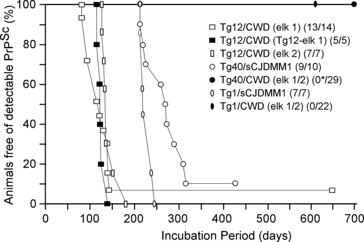
Chronic wasting disease (CWD), a prion disease affecting free-ranging and captive cervids (deer and elk), is widespread in the United States and parts of Canada. The large cervid population, the popularity of venison consumption, and the apparent spread of the CWD epidemic are likely resulting in increased human exposure to CWD in the United States. Whether CWD is transmissible to humans, as has been shown for bovine spongiform encephalopathy (the prion disease of cattle), is unknown. We generated transgenic mice expressing the elk or human prion protein (PrP) in a PrP-null background. After intracerebral inoculation with elk CWD prion, two lines of "humanized" transgenic mice that are susceptible to human prions failed to develop the hallmarks of prion diseases after >657 and >756 d, respectively, whereas the "cervidized" transgenic mice became infected after 118-142 d. These data indicate that there is a substantial species barrier for transmission of elk CWD to humans.


Figures

Figure 1.
Figure 1.
Survival curves of humanized and cervidized transgenic mice. Six- to eight-week-old cervidized Tg12 and humanized Tg1 and Tg40 mice were inoculated intracerebrally with 30 μl of 1% brain homogenate from two CWD-affected elk or a subject with sCJDMM1. The Tg12 mice were also inoculated with brain homogenates from elk 1 CWD-affected Tg12 mice to evaluate the species barrier of elk CWD transmission to the Tg12 mice. The average incubation times were as follows: 118±6 d for elk 1 CWD-inoculated Tg 12 mice (opensquares); 125±4 d for secondary transmission of elk 1 CWD in Tg12 mice (filled squares); 142 ± 7 d for elk 2 CWD-inoculated Tg12 mice (open rectangles); 263 ± 13 d for the sCJDMM1-inoculated Tg40 mice (open circles); and 226 ± 5 d for the sCJDMM1-inoculated Tg1 mice (open ovals). None of the 29 CWD-inoculated Tg40 mice (filled circle) or the 22 CWD-inoculated Tg1 mice (filled oval) had detectable PK-resistant PrPSc or substantial histopathology after >756 and >657 dpi, respectively. The asterisk indicates that three Tg40 mice became ataxic between 420 and 509 dpi but were free of prion infection. Parentheses indicate prion-positive mice per total inoculated mice.
Figure 2.
Figure 2.
Histopathology, PrP immunohistochemistry, and apoptosis examination in CWD-affected Tg12-cervidized mice. Prominent spongiform degeneration was present in the cerebral cortex (a) along with marked neuronal loss in the granule cell layer of the cerebellum (b) when compared with age-matched control Tg12 mice (c) (hematoxylin and eosin staining; 20× magnification). PrP deposits formed a fine granular pattern interspersed with plaque-like deposits (arrow) in the cerebral cortex (d) and in the granule cell layer of the cerebellum (e) (mAb 8H4; 20× magnification). Neuronal apoptosis (arrow) was present in the granule cells of the hippocampus (f) and in other brain regions (TUNEL; 40× magnification).
Figure 3.
Figure 3.
Characterization of PK-resistant PrPSc from the donor CWD-affected elk and CWD-affected Tg12 mice. a, Immunoblot of PrP. Lanes 1 and 2, PK-untreated (lane 1) and PK-treated (lane 2) PrP from one of the donor elks with CWD. Lanes 3–14, PK-untreated (lane 3) and PK-treated (lanes 4–14) PrP from 11 CWD-affected Tg12 mice. b, Glycoform ratio analysis of PK-resistant PrPSc. The blots were probed with mAb 8H4. Error bars are based on quantitative analyses of digital chemiluminescence images of triplicate Western blots of brain homogenates from an elk with CWD (the inoculum) and duplicate Western blots of 11 Tg12 mice infected with elk CWD. kD, Kilodalton.
Figure 4.
Figure 4.
Absence of PK-resistant PrPSc in elk CWD-inoculated humanized Tg mice. Brain homogenates were subjected to sodium phosphotungstate treatment to precipitate PrPSc, digested with 20 μg/ml PK for 30 min, and analyzed by Western blotting with the mAb 3F4. Lane 1, An uninoculated Tg40 mouse; lane 2, an sCJDMM1-infected Tg40 mouse; lanes 3–5, three ataxic CWD-inoculated Tg40 mice; lanes 6–8, three CWD-inoculated Tg40 mice killed because of other aging-related illnesses. Lanes 1, 3–8, Forty microliters of 10% brain homogenates were loaded; lane 2, a total of 2 μl was loaded. kD, Kilodalton.
Figure 5.
Figure 5.
Histopathology and PrPSc deposition in the brains of sCJDMM1-infected Tg40 mice. In the brains of sCJDMM1-infected Tg40 mice, hematoxylin and eosin staining revealed moderate spongiform degeneration with fine vacuoles in the cerebral cortex (a) accompanied by even milder spongiosis in other brain areas (20×magnification), but the granule cell layer of the cerebellum (b) appears mostly intact (40× magnification). Immunohistochemistry for PrPSc with 3F4 revealed fine PrPSc deposits in the cerebral cortex (c), with more intense PrPSc deposits in the cerebellum (d) (20× magnification).
Figure 6.
Figure 6.
Immunoblots and glycoform ratios of PK-resistant PrPSc from the sCJDMM1 donor and sCJDMM1-inoculated Tg1 mice. a, Immunoblot of PrP. PK-untreated (lane 1) PrP and PK-treated (lane 2) PrP were obtained from the sCJDMM1 donor; PK-untreated (lane 3) PrP and PK-treated (lanes 4–10) PrP were obtained from the seven sCJDMM1-inoculated Tg1 mice. b, Glycoform ratio analysis of PK-resistant PrPSc. PK-untreated and PK-treated brain homogenates were processed as in Figure 2, but blots were probed with 3F4. Error bars are based on quantitative analyses of digital chemiluminescence images of triplicate Western blots of brain homogenates from a subject with sCJDMM1 (the inoculum) and duplicate Western blots of the seven Tg1 mice infected with sCJDMM1. kD, Kilodalton.

References

    1. Asante EA, Linehan JM, Desbruslais M, Joiner S, Gowland I, Wood AL, Welch J, Hill AF, Lloyd SE, Wadsworth JD, Collinge J (2002) BSE prions propagate as either variant CJD-like or sporadic CJD-like prion strains in transgenic mice expressing human prion protein. EMBO J 21: 6358–6366. - PMC - PubMed
    1. Belay ED, Gambetti P, Schonberger LB, Parchi P, Lyon DR, Capellari S, McQuiston JH, Bradley K, Dowdle G, Crutcher JM, Nichol CR (2001) Creutzfeldt-Jakob disease in unusually young patients who consumed venison. Arch Neurol 58: 1673–1678. - PubMed
    1. Belay ED, Maddox RA, Gambetti P, Schonberger LB (2003) Monitoring the occurrence of emerging forms of Creutzfeldt-Jakob disease in the United States. Neurology 60: 176–181. - PubMed
    1. Belay ED, Maddox RA, Williams ES, Miller MW, Gambetti P, Schonberger LB (2004) Chronic wasting disease and potential transmission to humans. Emerg Infect Dis 10: 977–984. - PMC - PubMed
    1. Browning SR, Mason GL, Seward T, Green M, Eliason GA, Mathiason C, Miller MW, Williams ES, Hoover E, Telling GC (2004) Transmission of prions from mule deer and elk with chronic wasting disease to transgenic mice expressing cervid PrP. J Virol 78: 13345–13350. - PMC - PubMed

Publication types

LinkOut - more resources