. 2005 Sep;25(18):8166-78.
doi: 10.1128/MCB.25.18.8166-8178.2005.

Pocket protein complexes are recruited to distinct targets in quiescent and proliferating cells

Affiliations

Pocket protein complexes are recruited to distinct targets in quiescent and proliferating cells

Egle Balciunaite et al. Mol Cell Biol. 2005 Sep.

Abstract

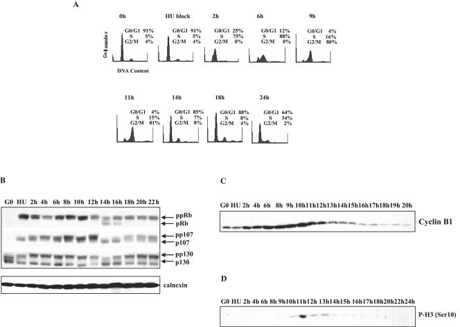
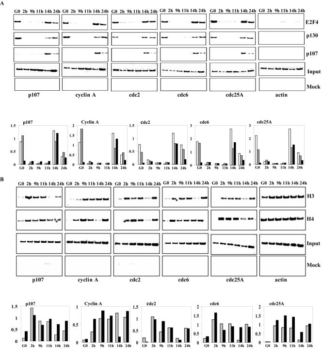
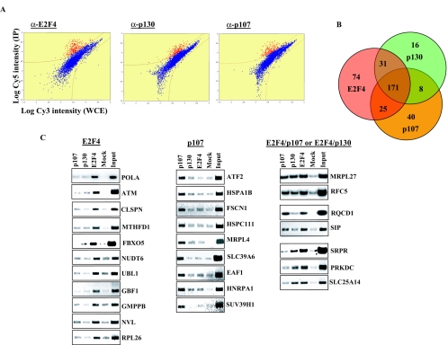
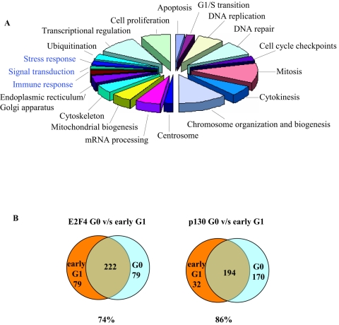
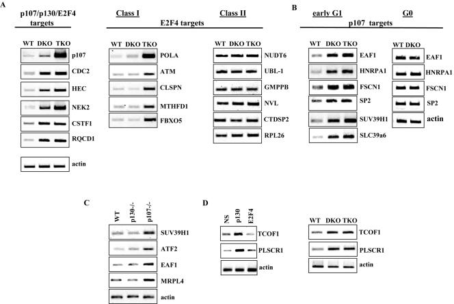
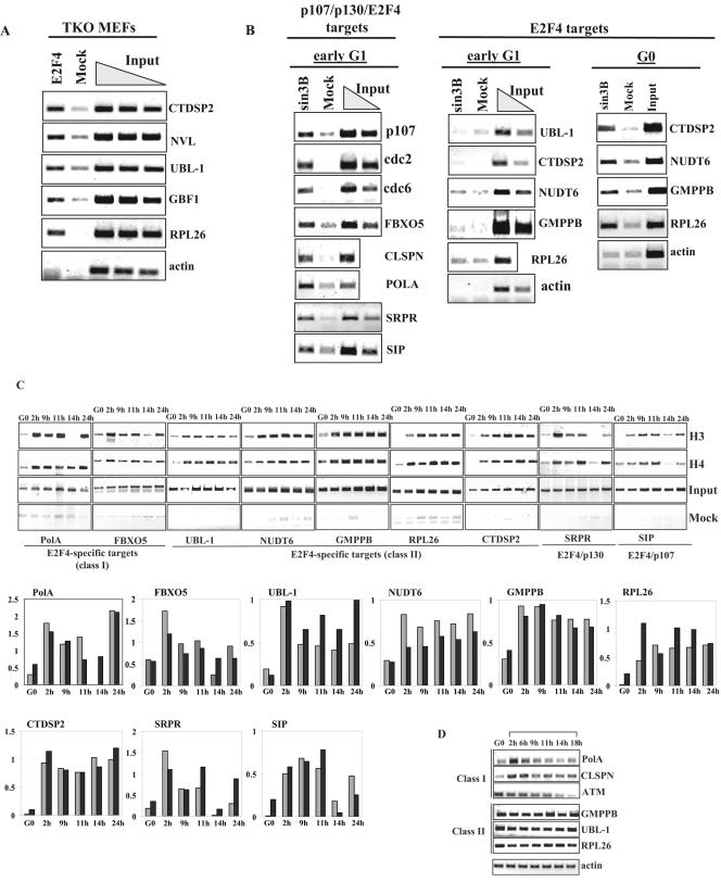
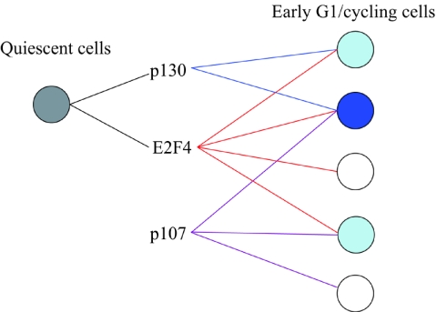
Biochemical and genetic studies have determined that retinoblastoma protein (pRB) tumor suppressor family members have overlapping functions. However, these studies have largely failed to distinguish functional differences between the highly related p107 and p130 proteins. Moreover, most studies pertaining to the pRB family and its principal target, the E2F transcription factor, have focused on cells that have reinitiated a cell cycle from quiescence, although recent studies suggest that cycling cells exhibit layers of regulation distinct from mitogenically stimulated cells. Using genome-wide chromatin immunoprecipitation, we show that there are distinct classes of genes directly regulated by unique combinations of E2F4, p107, and p130, including a group of genes specifically regulated in cycling cells. These groups exhibit both distinct histone acetylation signatures and patterns of mammalian Sin3B corepressor recruitment. Our findings suggest that cell cycle-dependent repression results from recruitment of an unexpected array of diverse complexes and reveals specific differences between transcriptional regulation in cycling and quiescent cells. In addition, factor location analyses have, for the first time, allowed the identification of novel and specific targets of the highly related transcriptional regulators p107 and p130, suggesting new and distinct regulatory networks engaged by each protein in continuously cycling cells.


Figures

FIG. 1.
FIG. 1.
Synchronization of T98G cells. (A) Cell synchronization. Quiescent T98G cells were stimulated with medium containing 10% FBS in the presence of 2 mM hydroxyurea (HU). After 22 h, the G1/S-arrested cells were washed and released into DMEM plus 10% FBS. At the indicated times following release from hydroxyurea, cells were harvested, stained with propidium iodide, and processed for FACS analysis. (B to D) T98G cells were synchronized as for panel A. At the indicated times following release from hydroxyurea, cell lysates were harvested and subjected to SDS-PAGE and Western blot analysis with antibodies against (B) Rb, p107, and p130, (C) cyclin B1, and (D) phosphorylated histone H3 (Ser 10). A protein abundance control (calnexin) was included as indicated.
FIG. 2.
FIG. 2.
Promoter occupancy and acetylation of histones H3 and H4 throughout the cell cycle. (A) T98 G cells were synchronized by hydroxyurea (HU) block. Cells were released for 2, 9, 11, 14, and 24 h to obtain enriched populations of cells in S, G2, M, early G1, and G1/S phases, respectively. Chromatin was prepared and immunoprecipitated with antibodies against E2F4, p107, p130, or p57 (negative control). The resulting ChIPs were amplified with primers to the promoters of p107, cyclin A, cdc2, cdc6, cdc25A, and actin. The input reaction represents 0.5% of the total chromatin for each sample. (B) T98G cells were treated as above and ChIP analysis was performed using antibodies against the panacetylated forms of histone H3 and histone H4. Histograms for both panels A and B show the relative binding activity of each transcription factor or the extent of acetylation of histones as quantitated from data in the upper panel. In panel A, white, gray, and black bars represent binding of E2F4, p130, and p107, respectively. In panel B gray and black bars represent acetylated H3 and acetylated H4, respectively.
FIG. 3.
FIG. 3.
Location analysis of E2F4, p107 and p130 in early G1 of cycling T98G cells. (A). Representative scatter plots for location analyses on Hu13K promoter microarrays. Fluorescence intensities in the Cy5 channel (ChIP) are plotted against the Cy3 channel (input chromatin). Red spots represent enriched promoters and blue spots represent nonenriched promoters. (B). Venn diagram comparing E2F4, p130, and p107 targets. (C). Subset of targets identified by ChIP-on-Chip confirmed by conventional ChIP and gene-specific PCR. Chromatin immunoprecipitation was performed as described in Materials and Methods. The input lane indicates PCRs containing 0.25% of total chromatin for each sample.
FIG. 4.
FIG. 4.
Functional categories of ChIP-on-chip targets in constantly cycling and growth inhibited cells. (A) Pie chart describing major functional gene ontology categories of E2F4, p107, and p130 targets enriched in the early G1 phase of cycling cells. Functional categories in blue represent targets identified in cycling cells but not growth-inhibited cells. (B) Venn diagrams comparing E2F4 and p130 targets in the early G1 phase of cycling cells to that of G0 phase in quiescent cells.
FIG. 5.
FIG. 5.
Regulation of E2F4-specific targets in early G1 cells deficient for p107/p130, and all pocket proteins. (A to D) RT-PCR analysis of targets identified by location analysis. (A) Expression of a subset of p107/p130/E2F4- and E2F4-specific targets in p107- and p130-null MEFs (DKO), and p107-, p130-, and pRB-null MEFs (TKO). Prior to RT-PCR analysis, MEFs were synchronized in early G1 by hydroxyurea block and release for 14 h. Class I E2F4 targets are those that show deregulation in triple-knockout but not double-knockout MEFs. Class II E2F4 targets are those that are not deregulated in either double-knockout or triple-knockout MEFs. (B) Expression of a subset of p107 targets in wild-type (WT), double-knockout (DKO), and triple-knockout (TKO) MEFs. Prior to RT-PCR analysis, MEFs were synchronized in either G0 or G1 as described in Materials and Methods. (C) Expression of p107 targets in p107- or p130-null MEFs. Cells were treated as for panel A. (D) Left panel: RT-PCR analysis of p130 targets in T98G (human) cells treated with nonspecific siRNA or siRNA specific for p130. T98G cell were synchronized by hydroxyurea (HU) treatment and released for 3 h from hydroxyurea block. Right panel: RT-PCR analysis of two p130 targets in early G1 double-knockout MEFs and triple-knockout MEFs.
FIG. 6.
FIG. 6.
Analysis of E2F-specific promoters. (A) ChIP of E2F4 in triple-knockout (TKO) MEFs. Various amounts of total chromatin (0.5, 0.25, and 0.125%) were used to estimate the relative binding of E2F4 to the promoters. (B) ChIP of Sin3B in T98G cells. Cells were synchronized by hydroxyurea (HU) block and released for 14 h to enrich for cells in early G1 phase or serum starved for 72 h (G0). (C) Acetylation of E2F4-specific promoters. T98G cells were treated as described in the legend to Fig. 2B. Gene names and classifications are indicated. Graphs for both panels show the relative extent of acetylation of histones as quantitated from data in the figure. Gray and black bars represent acetylated H3 and acetylated H4, respectively. (D) Expression analysis of E2F4-only class I and class II genes. T98G cells were treated with hydroxyurea and released from this block for the indicated times as described in the legend to Fig. 2B. The brackets indicate times after release from hydroxyurea.
FIG. 7.
FIG. 7.
Model summarizing different transcriptional networks regulated by E2F4 and/or pocket proteins in quiescent and early G1 cells. Color intensity indicates the number of factors that regulate a given class of target genes.

References

    1. Alland, L., R. Muhle, H. Hou, Jr., J. Potes, L. Chin, N. Schreiber-Agus, and R. A. DePinho. 1997. Role for N-CoR and histone deacetylase in Sin3-mediated transcriptional repression. Nature 387:49-55. - PubMed
    1. Bissell, M. J., and M. A. Labarge. 2005. Context, tissue plasticity, and cancer: are tumor stem cells also regulated by the microenvironment? Cancer Cell 7:17-23. - PMC - PubMed
    1. Blais, A., and B. D. Dynlacht. 2004. Hitting their targets: an emerging picture of E2F and cell cycle control. Curr. Opin. Genet. Dev. 14:527-532. - PubMed
    1. Blais, A., M. Tsikitis, D. Acosta-Alvear, R. Sharan, Y. Kluger, and B. D. Dynlacht. 2005. An initial blueprint for myogenic differentiation. Genes Dev. 19:533-569. - PMC - PubMed
    1. Cam, H., E. Balciunaite, A. Blais, A. Spektor, R. C. Scarpulla, R. Young, Y. Kluger, and B. D. Dynlacht. 2004. A common set of gene regulatory networks links metabolism and growth inhibition. Mol. Cell 16:399-411. - PubMed

Publication types

MeSH terms

LinkOut - more resources