. 2005 Sep 1;437(7055):94-100.
doi: 10.1038/nature04029.

Human subtelomeres are hot spots of interchromosomal recombination and segmental duplication

Affiliations

Human subtelomeres are hot spots of interchromosomal recombination and segmental duplication

Elena V Linardopoulou et al. Nature. .

Abstract

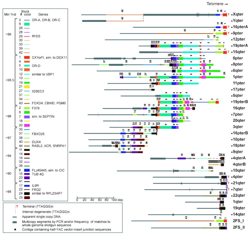
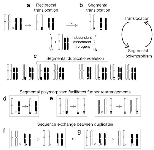
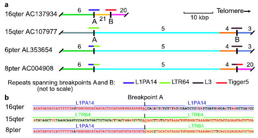
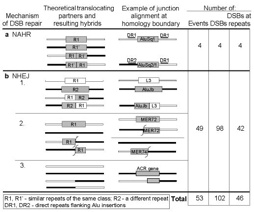
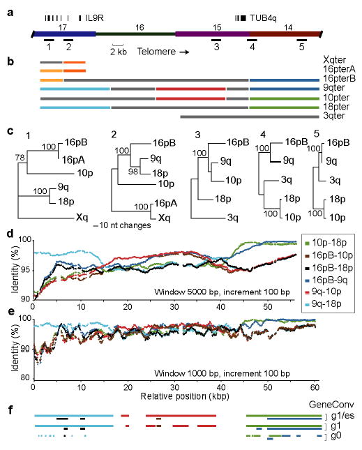
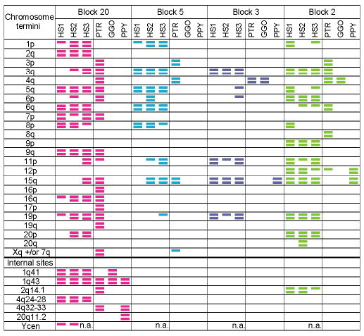
Human subtelomeres are polymorphic patchworks of interchromosomal segmental duplications at the ends of chromosomes. Here we provide evidence that these patchworks arose recently through repeated translocations between chromosome ends. We assess the relative contribution of the principal mechanisms of ectopic DNA repair to the formation of subtelomeric duplications and find that non-homologous end-joining predominates. Once subtelomeric duplications arise, they are prone to homology-based sequence transfers as shown by the incongruent phylogenetic relationships of neighbouring sections. Interchromosomal recombination of subtelomeres is a potent force for recent change. Cytogenetic and sequence analyses reveal that pieces of the subtelomeric patchwork have changed location and copy number with unprecedented frequency during primate evolution. Half of the known subtelomeric sequence has formed recently, through human-specific sequence transfers and duplications. Subtelomeric dynamics result in a gene duplication rate significantly higher than the genome average and could have both advantageous and pathological consequences in human biology. More generally, our analyses suggest an evolutionary cycle between segmental polymorphisms and genome rearrangements.


Figures

Figure 1
Figure 1
Subtelomeric paralogy map. Subtelomeric contigs (Table S1 gives constituent accessions and localization methods) are aligned at telomeres or to maximize alignments of paralogous blocks. Copies of a given block have the same color, width, and number. Only blocks 15 and 40 on 4q, 22 on 3q, 34–37 on 1p, and 38 on 6q are in inverted orientation relative to other corresponding block copies. 2qFS_I and _II represent ancestral telomeres fused head-to-head at 2q13–14; other interstitial paralogies are not displayed or analyzed here. A and B indicate allelic variants. Yq/Xq pseudoautosomal homology extends distal of dotted line.
Figure 2
Figure 2
A translocation-based model of segmental duplication and polymorphism. (a) A terminal duplication/deletion can arise if a translocation product and an intact homolog are passed from parent to offspring, creating a segmental polymorphism in (c). (b) A segmental duplication/deletion can arise if a second inter-chromosomal exchange occurs between the translocated chromosomes. Segmental polymorphism can facilitate further rearrangements by (d) promoting translocations through inter-chromosomal homologies, or (e) causing translocation or other rearrangement due to the absence of homology. Both reciprocal and non-reciprocal homology-based sequence transfers (f and g) are possible between duplicates generated by any of the above steps. *, sequence variant.
Figure 3
Figure 3
Layers of inter-chromosomal translocations form subtelomeric blocks. (a) Paralogous blocks have shared color and number; short colored lines above indicate different repetitive elements at homology breakpoints A and B, which define two translocations. An intact copy of each repeat is preserved in 16q and 15q sequences spanning the homology breakpoints with 6p and 8p, which contain truncated repeats fused by NHEJ. (b) Only two identical nucleotides (underlined) are found at the point where the original two sequences were joined at breakpoint A to form a hybrid. Aligned matching bases are red.
Figure 4
Figure 4
Most subtelomeric homology breakpoints are consistent with NHEJ. For each mechanistic scenario, we diagram both original and derived forms, assuming reciprocal exchange. One derived form would be lacking in non-reciprocal cases. The third column gives a schematic example of each scenario identified in pairwise alignments of subtelomeric homology blocks. Fifty-three of the complete, non-redundant set of 56 homology breakpoints were assigned a mechanistic scenario (details in Table S5, Fig. S3). In some cases, two originals and one hybrid were available for comparison (e.g., NHEJ group 1). Other predicted states were not among surviving, sequenced alleles.
Figure 5
Figure 5
Homology-based sequence transfers between subtelomeres. (a) The region analyzed encompasses four numbered blocks, two multi-exon genes, and five sequences sampled for phylogenetic analyses. (b) Diagram of multiple sequence alignment with colors (excluding gray) indicating the best matching pairs with ≥98% identity in non-overlapping 5-kbp windows. (c) Neighbor-joining trees with bootstrap values (over 1000 replicates) constructed from 2-kbp samples of the alignments. (d) and (e) Plot of percent identity between four subtelomeres in 5-kbp and 1-kbp windows, respectively. Colors indicate alignments of different pairs. (f) The same colors indicate transferred segments found statistically significant by GeneConv with different stringency parameters.
Figure 6
Figure 6
Chromosomal distribution of four subtelomeric blocks. FISH was conducted on three unrelated humans (HS1-3), chimpanzee (PTR), gorilla (GGO), and orangutan (PPY) (see Supplementary Methods). Colored bars indicate sites at which FISH signals were consistently observed on both homologs (two bars) or only one homolog (one bar). Colors correspond to Fig. 1. Chromosome locations are given according to the human karyotype. No signal was observed for block 5 in gorilla and orangutan; its presence was also not detected by PCR (Table S3).

References

    1. Samonte RV, Eichler EE. Segmental duplications and the evolution of the primate genome. Nat Rev Genet. 2002;3:65–72. - PubMed
    1. Bailey JA, et al. Recent segmental duplications in the human genome. Science. 2002;297:1003–7. - PubMed
    1. Bailey JA, Yavor AM, Massa HF, Trask BJ, Eichler EE. Segmental duplications: organization and impact within the current human genome project assembly. Genome Res. 2001;11:1005–17. - PMC - PubMed
    1. Shaw CJ, Lupski JR. Implications of human genome architecture for rearrangement-based disorders: the genomic basis of disease. Hum Mol Genet. 2004;13(Spec No 1):R57–64. - PubMed
    1. Mefford H, Trask BJ. The complex structure and dynamic evolution of human subtelomeres. Nature Reviews Genetics. 2002;3:91–102. - PubMed

Publication types