. 2005 Sep 1;437(7055):154-8.
doi: 10.1038/nature03923.

Structural mechanism for sterol sensing and transport by OSBP-related proteins

Affiliations

Structural mechanism for sterol sensing and transport by OSBP-related proteins

Young Jun Im et al. Nature. .

Abstract

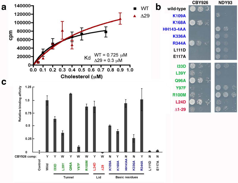
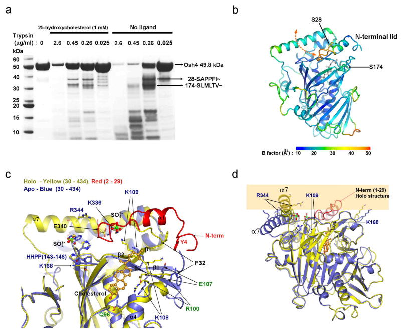
The oxysterol-binding-protein (OSBP)-related proteins (ORPs) are conserved from yeast to humans, and are implicated in the regulation of sterol homeostasis and in signal transduction pathways. Here we report the structure of the full-length yeast ORP Osh4 (also known as Kes1) at 1.5-1.9 A resolution in complexes with ergosterol, cholesterol, and 7-, 20- and 25-hydroxycholesterol. We find that a single sterol molecule binds within a hydrophobic tunnel in a manner consistent with a transport function for ORPs. The entrance is blocked by a flexible amino-terminal lid and surrounded by basic residues that are critical for Osh4 function. The structure of the open state of a lid-truncated form of Osh4 was determined at 2.5 A resolution. Structural analysis and limited proteolysis show that sterol binding closes the lid and stabilizes a conformation favouring transport across aqueous barriers and signal transmission. The structure of Osh4 in the absence of ligand exposes potential phospholipid-binding sites that are positioned for membrane docking and sterol exchange. On the basis of these observations, we propose a model in which sterol and membrane binding promote reciprocal conformational changes that facilitate a sterol transfer and signalling cycle.


Figures

Figure 1
Figure 1. Structure of Osh4
a. The overall structure of Osh4. The N-terminal lid (1–29) is red, the central helices (30–116) orange, the β-barrel (117–307) green, and the C-terminal sub-domain (308–434) cyan. b. Surface Osh4: basic residues are blue, acidic red, hydrophobic green, and neutral polar white. Waters are shown as red spheres. c. Strictly conserved residues are colored cyan. The N-terminal lid is shown in ribbons and colored red. d. Solvent-accessible conserved residues are clustered around the tunnel entrance. e. Residues that bind 25-HC are colored green. f. Recognition of 25-HC 25-hydroxyl group. The ɛ-amino group of Lys336 has two conformations in all complex crystal structures, with the left-hand conformation (closest to Lys 109) predominating. g. Superposition of five sterols in the binding site. 7-HC is colored gray, 20-HC cyan, 25-HC red, cholesterol green and ergosterol blue. Hydroxyl groups in the sterols are shown in spheres. Hydrogen bonds are shown in dashed lines.
Figure 2
Figure 2. Mutational analysis of binding and function
a. Binding curves for wild-type and lid truncation mutant using low-stringency washing protocol. Each data point shown is the average of two measurements. b. Plasmids encoding Osh4 mutants were introduced into CBY926 (4) and NDY93. The strains were grown at permissive temperature (23°C) and dilution series were incubated at 37°C. All experiments were repeated at least 3 times with the same results. Expression of mutants in vivo was quantitated by immunoblotting (Supplementary Fig. 6). c. CPM measured for labeled cholesterol bound at a concentration of 0.15 μM to Osh4 mutants were scaled to wild type (100 %) using high-stringency washing protocol. The standard error from three measurements is shown by the bars.
Figure 3
Figure 3. Conformational changes in Osh4
a. Tryptic digestion of Osh4. The arrows indicate the N-terminal amino acid sequences of the bands. b. The ribbon model is colored by the average B-factor of the residue to show their relative mobility. Trypsin cleavage sites are indicated by arrows. c. Apo (blue) and cholesterol complex (red for lid region and yellow elsewhere) structures were superimposed. The sulfate ions bound to the apo structure in positions that membrane phospholipid phosphates are shown. d. Superposition of apo and cholesterol complex structures, with the location of the membrane indicated by yellow shading.
Figure 4
Figure 4. Mechanism of sterol transfer
The proposed cholesterol transfer cycle is depicted. Oxysterols are shown binding via the cytosol rather than the membrane because of their higher solubility. Cholesterol and oxysterol-dependent signals are shown as occurring via the bound conformation in the cytosol as found for OSBP.

Comment in

Similar articles

Cited by

References

    1. Dawson PA, Ridgway ND, Slaughter CA, Brown MS, Goldstein JL. cDNA Cloning and Expression of Oxysterol-Binding Protein, an Oligomer with a Potential Leucine Zipper. J Biol Chem. 1989;264:16798–16803. - PubMed
    1. Olkkonen VM, Levine TP. Oxysterol binding proteins: in more than one place at one time? Biochem Cell Biol. 2004;82:87–98. - PubMed
    1. Beh CT, Cool L, Phillips J, Rine J. Overlapping functions of the yeast oxysterol-binding protein homologues. Genetics. 2001;157:1117–1140. - PMC - PubMed
    1. Beh CT, Rine J. A role for yeast oxysterol-binding protein homologs in endocytosis and in the maintenance of intracellular sterol-lipid distribution. J Cell Sci. 2004;117:2983–2996. - PubMed
    1. Wang P, Weng J, Anderson RGW. OSBP is a cholesterol-regulated scaffolding protein in control of ERK1/2 activation. Science. 2005;307:1472–1476. - PubMed

Publication types

MeSH terms