

What parameters affect left ventricular diastolic flow propagation velocity? In vitro studies using color M-mode Doppler echocardiography
- PMID: 16137332
- PMCID: PMC1236942
- DOI: 10.1186/1476-7120-3-24
What parameters affect left ventricular diastolic flow propagation velocity? In vitro studies using color M-mode Doppler echocardiography
Abstract
Background: Insufficient data describe the relationship of hemodynamic parameters to left ventricular (LV) diastolic flow propagation velocity (Vp) measured using color M-mode Doppler echocardiography.
Methods: An in vitro LV model used to simulate LV diastolic inflow with Vp measured under conditions of varying: 1) Stroke volume, 2) heart rate (HR), 3) LV volume, 4) LV compliance, and 5) transmitral flow (TMF) waveforms (Type 1: constant low diastasis flow and Type 2: no diastasis flow).
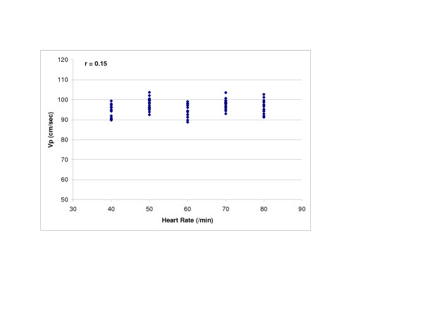
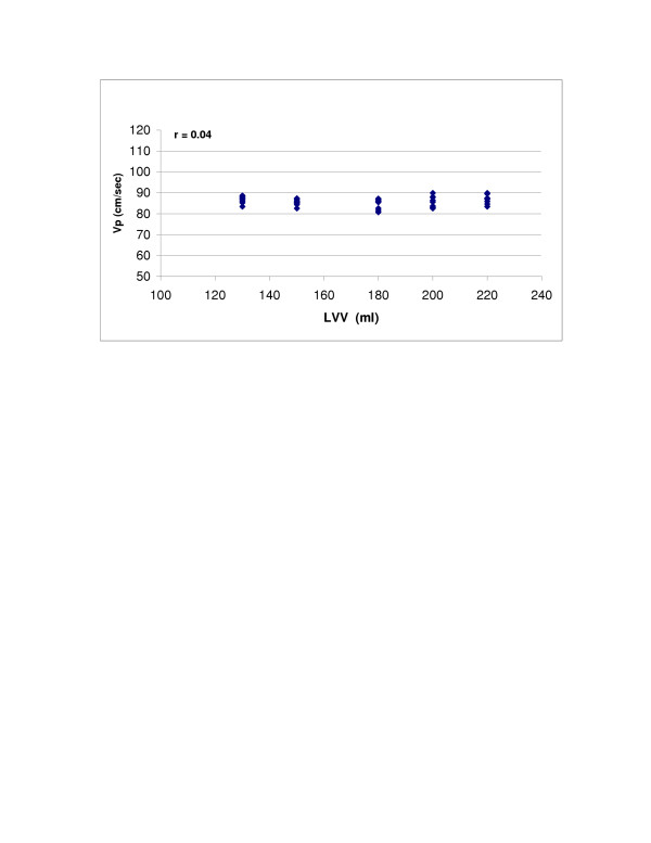
Results: Univariate analysis revealed excellent correlations of Vp with stroke volume (r = 0.98), LV compliance (r = 0.94), and HR with Type 1 TMF (r = 0.97). However, with Type 2 TMF, HR was not associated with Vp. LV volume was not related to Vp under low compliance, but inversely related to Vp under high compliance conditions (r = -0.56).
Conclusion: These in vitro findings may help elucidate the relationship of hemodynamic parameters to early diastolic LV filling.
Figures


Similar articles
-
Assessment of propagation velocity by contrast echocardiography for standardization of color Doppler propagation velocity measurements.J Am Soc Echocardiogr. 2004 Dec;17(12):1266-74. doi: 10.1016/j.echo.2004.08.028. J Am Soc Echocardiogr. 2004. PMID: 15562265
-
Effect of heart rate on tissue Doppler measures of diastolic function.Echocardiography. 2007 Aug;24(7):697-701. doi: 10.1111/j.1540-8175.2007.00466.x. Echocardiography. 2007. PMID: 17651098
-
A new Doppler method for assessing left ventricular diastolic stiffness based on principles of flow wave propagation: mathematical basis and review of the method.J Heart Valve Dis. 1993 Mar;2(2):167-73. J Heart Valve Dis. 1993. PMID: 8261154
-
[Echocardiographic and Doppler echocardiographic characterization of left ventricular diastolic function].Herz. 1990 Dec;15(6):377-92. Herz. 1990. PMID: 2279732 Review. German.
-
Assessment of left ventricular dimensions and functions in athletes and sedentary subjects at rest and during exercise using echocardiography, Doppler sonography and radionuclide ventriculography.Int J Sports Med. 1996 Nov;17 Suppl 3:S173-9. doi: 10.1055/s-2007-972920. Int J Sports Med. 1996. PMID: 9119539 Review.
Cited by
-
Beta-adrenergic receptor desensitization in man: insight into post-exercise attenuation of cardiac function.J Physiol. 2006 Dec 1;577(Pt 2):717-25. doi: 10.1113/jphysiol.2006.116426. Epub 2006 Sep 14. J Physiol. 2006. PMID: 16973702 Free PMC article.
-
Anthrax toxins induce shock in rats by depressed cardiac ventricular function.PLoS One. 2007 May 23;2(5):e466. doi: 10.1371/journal.pone.0000466. PLoS One. 2007. PMID: 17520025 Free PMC article.
References
-
- Brun P, Tribouilloy C, Duval AM, Iseriu L, Megurira A, Pelle G, Dubois-Rande JL. Left ventricular flow propagation during early filling is related to wall relaxation: a color M-mode Doppler analysis. J Am Coll Cardiol. 1992;20:420–432. - PubMed
-
- Stugaard M, Risoe C, Halfdan I, Smiseth OA. Intraventricular early diastolic filling during acute myocardial ischemia: assessment by multigated color M-mode Doppler echocardiography. Circulation. 1993;88:2705–2713. - PubMed
-
- Takatsuji H, Mikami T, Urasawa K, Teranishi J-I, Onozuka H, Takagi C, Makita Y, Matsuo H, Kusuoka H, Kitabatake A. A new approach for evaluation of left ventricular diastolic function: Spatial and temporal analysis of left ventricular filling flow propagation by color M-mode Doppler echocardiography. J Am Coll Cardiol. 1996;27:365–371. doi: 10.1016/0735-1097(96)81240-X. - DOI - PubMed
-
- Garcia MJ, Smedira NG, Greenberg NL, Main M, Firstenberg MS, Odabashian J, Thomas JD. Color M-mode Doppler flow propagation velocity is a preload insensitive index of left ventricular relaxation: animal and human validation. J Am Coll Cardiol. 2000;35:201–208. doi: 10.1016/S0735-1097(99)00503-3. - DOI - PubMed
-
- Yellin EL, Hori M, Yorcan C, Sonnenblick EH, Gabbay S, Frater RW. Left ventricular relaxation in the filling and nonfilling intact canine heart. Am J Physiol. 1986;250:H620–629. - PubMed
MeSH terms
LinkOut - more resources
Full Text Sources
