. 2005 Oct;20(1):64-73.
doi: 10.1016/j.nbd.2005.02.001.

Tau gene transfer, but not alpha-synuclein, induces both progressive dopamine neuron degeneration and rotational behavior in the rat

Affiliations

Tau gene transfer, but not alpha-synuclein, induces both progressive dopamine neuron degeneration and rotational behavior in the rat

Ronald L Klein et al. Neurobiol Dis. 2005 Oct.

Abstract

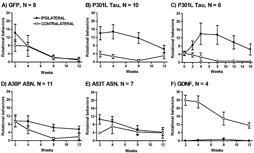
Using a viral vector for mutant (P301L) tau, we studied the effects of gene transfer to the rat substantia nigra in terms of structural and functional properties of dopaminergic neurons. The mutant tau vector caused progressive loss of pars compacta dopaminergic neurons over time, reduced striatal dopamine content, and amphetamine-stimulated rotational behavior consistent with a specific lesion effect. In addition, structural studies demonstrated neurofibrillary tangles and neuritic pathology. Wild-type tau had similar effects on neuronal loss and rotational behavior. In contrast, mutant alpha-synuclein vectors did not induce rotational behavior, although alpha-synuclein filaments formed in nigrostriatal axons. Dopamine neuron function is affected by tau gene transfer and appears to be more susceptible to tau- rather than alpha-synuclein-related damage in this model. Both tau and alpha-synuclein are important for substantia nigra neurodegeneration models in rats, further indicating their potential as therapeutic targets for human diseases involving loss of dopamine neurons.


Figures

Fig. 1
Fig. 1
Amphetamine-stimulated rotational behavior time-course. (A) Control GFP vector group 2–12 weeks after gene transfer. (B) P301L tau vector group 2– 12 weeks. (C) P301L tau vector group 0–16 weeks, separate group of rats from B. (D) A30P ASN vector group 2–12 weeks. (E) A53T ASN vector group 2–12 weeks. (F) GDNF vector group 2–12 weeks. The N values are shown on graphs. Repeated-measures ANOVA analyses described in Results. Directional differences found in the tau and GDNF vector groups (B, C, F; P < 0.01).
Fig. 2
Fig. 2
Western blots of dissected substantia nigra (SN) and striatum (STR) showing GFP, tau, or ASN expression from the respective vector. (A) GFP immunoblot, after injecting 2 × 10¹⁰ particles of the GFP AAV unilaterally to the rat SN. Band ~31 kDa, as expected. Lane 1, uninjected SN; lane 2, injected SN; lane 3, STR uninjected side; lane 4, STR injected side. (B) Human tau immunoblot, after injecting 2 × 10¹⁰ particles of the human P301L tau AAV unilaterally to the SN. Band ~70 kDa, as expected for the longest form of tau. Lane 1, uninjected SN; lane 2, injected SN; lane 3, uninjected SN from a second rat; lane 4, injected SN from second rat. We did not detect human tau in samples from STR. (C) ASN immunoblot, after injecting 2 × 10¹⁰ particles of the A30P ASN-WPRE AAV unilaterally to the SN. Lane 1, uninjected SN; lane 2, injected SN; lane 3, STR uninjected side; lane 4, STR injected side. (A–C) 4 months after gene transfer. 60 µg protein loaded in each lane.
Fig. 3
Fig. 3
Tau gene transfer results in the degeneration of the SN pars compacta. (A) GFP vector injections to the midbrain led to robust expression of GFP immunoreactivity in the SN pars compacta (SNc), the SN pars reticulata below (SNr), and above the SN along the needle track. (B) TH staining on the side of the GFP expression (left side of panel) was similar relative to the contralateral uninjected side. (C) Human-specific tau labeling after injection of the P301L human tau vector, showing characteristic blank area in the SN pars compacta. (D) Consistent with the human tau expression pattern, TH staining in the SN pars compacta was dramatically reduced after tau vector injections (left side of panel). (E) Human-specific ASN labeling after injection of the A30P human ASN vector. Unlike with tau gene transfer, there were many ASN-expressing neurons in the SN pars compacta in the ASN groups. (F) Reduced TH staining was associated with ASN vector injections (left side of panel) as we observed with a previous, less efficient version of the ASN vector (Klein et al., 2002a). The loss of staining with the P301L tau AAV was generally more intense and more consistent than observed with ASN vectors in this and the previous study. Staining controls in untransfected tissues were blank for GFP or human tau or ASN. (A–F) 4 months after gene transfer. Scale bars, A = 320 µm; B = 700 µm; C = 125 µm. C and E, and B, D, and F, are of the same magnification.
Fig. 4
Fig. 4
Neurofibrillary pathology in SN neurons detected with antibody Ab39. (A) Neuronal cell bodies and processes in the lateral SN expressing Ab39 immunoreactivity, 4 months post-injection of the P301L tau vector. (B) Ab39 labeling was prevalent in axonal spheroids in the medially-projecting axons in the P301L tau vector group, 3 weeks post-injection. (C) Ab39 labeling and axonal spheroids also found in the wild-type tau vector group at 3 weeks. Ab39 labeling found only in tau vector groups. Scale bar = 32 µm. A–C, same magnification.
Fig. 5
Fig. 5
Ultrastructural detection of tau or ASN filaments in the rat SN 4 months after gene transfer. (A) A SN neuron showing eccentric nucleus, pushed by densely packed filaments after P301L tau gene transfer. There is extensive deposition of 10-nm gold particles with immunoEM for hyperphosphorylated tau. (B) Higher magnification of straight ~15-nm tau filaments within the same neuron as A. (C) A myelinated axon in the substantia nigra containing ASN filaments. Filaments labeled with gold particles (10 nm) were found after A30P ASN gene transfer using the human-specific ASN antibody. The filaments did not co-label with antibodies for neurofilaments and were proximal to mitochondria (M). (D) Higher magnification of a field in C. CP13 (A and B) or LB509 (C and D) antibody labeling was not observed in controls. Scale bars, A = 1 µm; B = 180 nm; C = 300 nm; D = 60 nm.

References

    1. Allen B, Ingram E, Takao M, Smith MJ, Jakes R, Virdee K, Yoshida H, Holzer M, Craxton M, Emson PC, Atzori C, Migheli A, Crowther RA, Ghetti B, Spillantini MG, Goedert M. Abundant tau filaments and nonapoptotic neurodegeneration in transgenic mice expressing human P301S tau protein. J. Neurosci. 2002;22:9340–9351. - PMC - PubMed
    1. Andorfer C, Kress Y, Espinoza M, de Silva R, Tucker KL, Barde YA, Duff K, Davies P. Hyperphosphorylation and aggregation of tau in mice expressing normal human tau isoforms. J. Neurochem. 2003;86:582–890. - PubMed
    1. Baker M, Litvan I, Houlden H, Adamson J, Dickson D, Perez-Tur J, Hardy J, Lynch T, Bigio E, Hutton M. Association of an extended haplotype in the tau gene with progressive supranuclear palsy. Hum. Mol. Genet. 1999;8:711–715. - PubMed
    1. Bancher C, Brunner C, Lassmann H, Budka H, Jellinger K, Seitelberger F, Grundke-Iqbal I, Iqbal K, Wisniewski HM. Tau and ubiquitin immunoreactivity at different stages of formation of Alzheimer neurofibrillary tangles. Prog. Clin. Biol. Res. 1989;317:837–848. - PubMed
    1. Braak E, Sandmann-Keil D, Rub U, Gai WP, de Vos RA, Steur EN, Arai K, Braak H. Alpha-synuclein immunopositive Parkinson’s disease-related inclusion bodies in lower brain stem nuclei. Acta Neuropathol. (Berl) 2001;101:195–201. - PubMed

Publication types

MeSH terms

LinkOut - more resources