. 2005 Oct;6(10):866-79.
doi: 10.1111/j.1600-0854.2005.00322.x.

Intragranular vesiculotubular compartments are involved in piecemeal degranulation by activated human eosinophils

Affiliations

Intragranular vesiculotubular compartments are involved in piecemeal degranulation by activated human eosinophils

Rossana C N Melo et al. Traffic. 2005 Oct.

Abstract

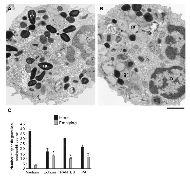
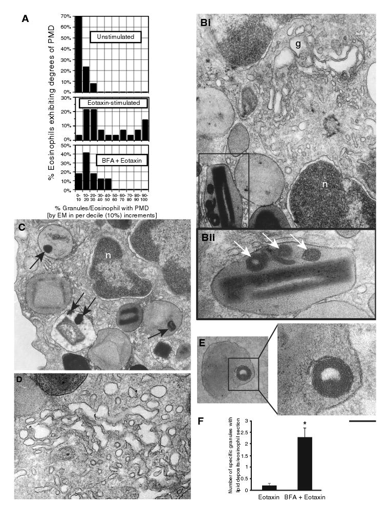
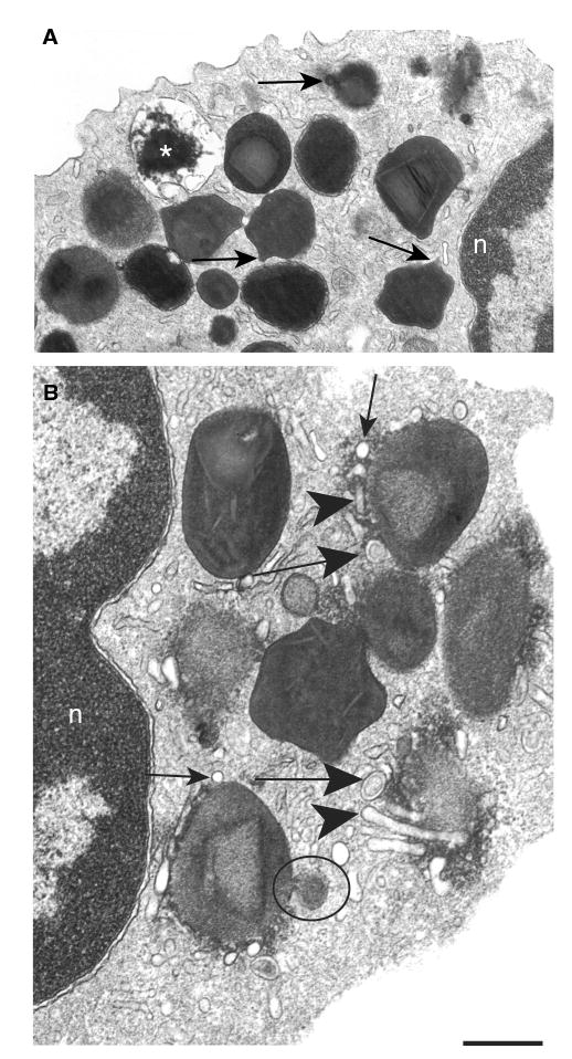
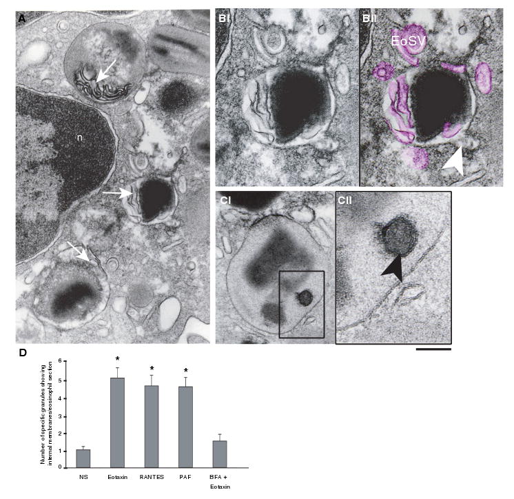
Eosinophils, leukocytes involved in allergic, inflammatory and immunoregulatory responses, have a distinct capacity to rapidly secrete preformed granule-stored proteins through piecemeal degranulation (PMD), a secretion process based on vesicular transport of proteins from within granules for extracellular release. Eosinophil-specific granules contain cytokines and cationic proteins, such as major basic protein (MBP). We evaluated structural mechanisms responsible for mobilizing proteins from within eosinophil granules. Human eosinophils stimulated for 30-60 min with eotaxin, regulated on activation, normal, T-cell expressed and secreted (RANTES) or platelet activating factor exhibited ultrastructural features of PMD (e.g. losses of granule contents) and extensive vesiculotubular networks within emptying granules. Brefeldin A inhibited granule emptying and collapsed intragranular vesiculotubular networks. By immunonanogold ultrastructural labelings, CD63, a tetraspanin membrane protein, was localized within granules and on vesicles outside of granules, and mobilization of MBP into vesicles within and extending from granules was demonstrated. Electron tomography with three dimension reconstructions revealed granule internal membranes to constitute an elaborate tubular network able to sequester and relocate granule products upon stimulation. We provide new insights into PMD and identify eosinophil specific granules as organelles whose internal tubulovesicular networks are important for the capacity of eosinophils to secrete, by vesicular transport, their content of preformed and granule-stored cytokines and cationic proteins.


Figures

Figure 1
Figure 1. Morphologic effect of three physiologic stimuli on human eosinophil specific granules
Cells were incubated with control buffer (A, C), 100 ng/mL eotaxin (B, C), 1 μM platelet-activating factor (PAF) (C) or 100 ng/mL regulated on activation, normal, T-cell expressed and secreted (RANTES) (C), immediately fixed and prepared for transmission electron microscopy. After 1 h of stimulation, granules exhibited progressive emptying of their contents and showed morphological diversity. Not all granules exhibited content losses. (C) Significant increases in numbers of emptying granules occurred after stimulation with the three stimuli (*p < 0.05). Eosinophils were isolated by negative selection from healthy donors. Counts were derived from three experiments with a total of 3945 granules counted in 95 electron micrographs randomly taken and showing the entire cell profile and nucleus. gr, granule; n, nucleus. Scale bar, 1.9 μm (A, B).
Figure 2
Figure 2. Morphology of emptying specific granules from stimulated human eosinophils
Granules exhibited lucent areas in their cores, matrices or both (A–D); reduced electron density (A, *); disassembled matrices and cores (A–D); residual cores (A, arrowheads) or membrane empty chambers (A, **). Intact, non-emptying granules (typical morphology B, *) were seen close to emptying granules. In (B and D), the arrows point to sites of vesicle budding profiles. A round small vesicle and large, vesiculotubular structures termed Eosinophil Sombrero Vesicles (EoSVs) are indicated by arrowheads, respectively, in (C and D). In (C), round profiles (circles) are within, and tubules surround (thin arrows) an emptying granule. Over 450 electron micrographs, ranging from ×10 000 to ×75 000 and taken from three different experiments, were evaluated. Eosinophils were isolated and stimulated for 1 h with eotaxin (A), platelet activating factor (B, D) or regulated on activation, normal, T-cell expressed and secreted (RANTES) (C) as in Figure 1, fixed and processed for transmission electron microscopy. n, nucleus. Scale bar, 630 nm (A) and 300 nm (B–D).
Figure 3
Figure 3. Brefeldin A (BFA) effects on eosinophil specific granules
(A) Heterogeneity of granule responses in unstimulated, eotaxin-stimulated and eotaxin plus BFA-pretreated (1 μg/mL, 30 min prior to eotaxin) eosinophils. Brefeldin A elicited the formation of membrano-lipid deposits within secretory granules (Bii and C, arrows. Bii corresponds to the boxed area in Bi), imaged as collapsed membranous structures (E, boxed area) and, as expected, disruption of the Golgi complex (Bi, D). (F) BFA induced significant increases in granule numbers showing internal lipid deposits (*p < 0.05). Granules with lipid deposits were numerated in a total of 50 electron micrographs randomly taken and showing the entire cell profile and nucleus. g, Golgi complex; n, nucleus; PMD, piecemeal degranulation. Scale bar, 650 nm (Bi), 208 nm (Bii), 630 nm (C), 500 nm (D), 340 nm (E) and 140 nm (E, higher magnification).
Figure 4
Figure 4. Early stimulus-induced granule events in human eosinophils
(A) After 30 min of stimulation, specific granules showed ill-defined cores and matrices and became irregular with elongation of their surfaces (arrows). An emptying granule is indicated (*). (B) A prominent system of vesicular compartments composed of small round vesicles (thin arrows), Eosinophil Sombrero Vesicles (EoSVs) (large arrows) and tubules (arrowheads) was seen at the granule surface. A large vesicle profile budding from the granule surface shows the same granule density (circle). Eosinophils were isolated and stimulated with eotaxin as in Figure 1. Transmission electron microscopic evaluations were derived from three experiments, and specimens were studied at magnifications ranging from ×5000 to ×75 000. n, nucleus. Scale bar, 720 nm (A) and 400 nm (B).
Figure 5
Figure 5. Vesicular transport of major basic protein (MBP) from within eotaxin-stimulated human eosinophils
Immunonanonogold electron microscopy revealed MBP-positive vesicles surrounding (A, C, E) and apparently arising (C, D) from mobilized specific granules. Control cells were negative for MBP labeling (B). In (Di), MBP labeling is seen on the disarranged granule core and matrix and within a small, spherical vesicle attached to the granule surface (box). Intragranular vesicles were also labeled for MBP. (Dii) corresponds to the boxed area of (Di). In (Ei), an eosinophil section shows the Golgi (G) region negative for MBP and MBP labeling within mobilized granules. MBP-positive, granule-associated large tubular carriers are indicated in (Eii) (arrowheads). (Eii) corresponds to the boxed area of (Ei). Cells were processed for pre-embedding immunonanogold labeling with mouse anti-MBP monoclonal antibody followed by 1.4 nm gold-conjugated goat-anti-mouse Fab fragments. In control cells, the primary antibody was replaced by an irrelevant antibody. In all experiments, eosinophils were stimulated as described in Figure 1. n, nucleus; gr, granule. Scale bar, 600 nm (A, B), 270 nm (C), 250 nm (Di), 150 nm (Dii), 500 nm (Ei) and 220 nm (Eii).
Figure 6
Figure 6. Emptying granules exhibit internal membranous domains in agonist-stimulated eosinophils
(A) Transmission electron microscopy (TEM) revealed intragranular membranous subcompartments organized as a tubular system in the matrix area (arrows). (B) Higher magnification of an emptying granule with internal membranous tubules (highlighted in pink in Bii) seen in conjunction with mobilized core. Eosinophil Sombrero Vesicles (EoSVs) around the granule are also colored pink. A budding vesicle is indicated (arrowhead). (Ci) A mobilized granule shows part of its content inside a subcompartment (box). (Cii) Higher magnification of the boxed area shows that a true internal membrane (arrowhead), with the same trilaminar appearance as exhibited by the outer granule membrane, encircles part of the granule content. (D) The numbers of granules showing internal membrane domains significantly increased after stimulation (*p < 0.05), but in brefeldin A (BFA)-pretreated eosinophils, these numbers did not differ from unstimulated cells. Cells were stimulated with eotaxin (A, B), regulated on activation, normal, T-cell expressed and secreted (RANTES) (C, D), platelet-activating factor (PAF) (D) or BFA prior to eotaxin (D), processed for TEM and analyzed as in Figure 1. n, nucleus. Scale bar, 480 nm (A), 300 nm (B), 350 nm (Ci) and 138 nm (Cii).
Figure 7
Figure 7. CD63 is localized to membranes of eosinophil specific granules
Phase-contrast and fluorescence microscopy of identical fields of a representative eotaxin-stimulated (A) and unstimulated (B) eosinophil. Both stimulated and unstimulated eosinophils exhibited fluorescent immunoreactive staining for CD63 on specific granules. In (C), deconvolution microscopy revealed peripheral CD63 labeling on specific granules from an unstimulated eosinophil. (D, E). Immunonanogold electron microscopy revealed CD63 labeling on the granule outer membranes (D) and within granule matrices (D, E, circles). (F) CD63-positive cytoplasmic vesicles showing membrane-bound labeling are indicated (arrows). (G) A control eosinophil showing negative labeling for CD63. Cells were stimulated and processed for immunofluorescence or pre-embedding immunogold labeling with anti-CD63 monoclonal antibody followed by, respectively, Alexa-488 (A), Alexa-594 (B) or 1.4 nm gold-conjugated (D–G) antibodies. In control cells, the primary antibody was replaced by an irrelevant antibody. Eosinophils were isolated and stimulated as Figure 1. n, nucleus. gr, granule. Scale bar, 6 μm (A, B), 5 μm (C), 350 nm (D), 400 nm (E, F) and 600 nm (G).
Figure 8
Figure 8. Tomographic slices and three dimension (3D) models from an emptying specific granule
(A–F) The tomographic volume shows intragranular subcompartments in conjunction with mobilized content. Circles indicate the same subcompartment surrounding part of the electron-dense content that is relocated to the granule outer membrane. Note in (C and D) that the electron density of this membrane changes at the site of contact with the membranous intragranular subcompartment. The arrows point to a forming Eosinophil Sombrero Vesicle (EoSV). Seventy and five serial single virtual slices as in (F) were extracted from the tomogram, outer granule membrane was partially traced in red and intragranular vesiculotubular structures were outlined in blue as in (G) so as to generate 3D models. (H–K) 3D models of the same granule show intragranular membrane domains (blue) organized as a flattened tubular network and tubules. In (J and K), the model has been rotated to provide another view. An area of continuity between the intragranular membranous network and the limiting granule membrane is indicated in (J, arrows). The slices (∼4 nm of thickness) were extracted from 3D reconstructions of a 400 nm eosinophil section analyzed by automated electron tomography at 200 kV. The numbers on the upper left corner indicate the slice number through the tomographic volume. Cells were stimulated with eotaxin as in Figure 1, chemically fixed and processed for transmission electron microscopy. Scale bar, 500 nm (A–F), 450 nm (G), 400 nm (H), 180 nm (I) and 150 nm (J, K). Also see Movie 1 and Movie 2 in the movie gallery gr, granule.

References

    1. Gleich GJ. Mechanisms of eosinophil-associated inflammation. J Allergy Clin Immunol. 2000;105:651–663. - PubMed
    1. Humbles AA, Lloyd CM, McMillan SJ, Friend DS, Xanthou G, McKenna EE, Ghiran S, Gerard NP, Yu C, Orkin SH, Gerard C. A critical role for eosinophils in allergic airways remodeling. Science. 2004;305:1776–1779. - PubMed
    1. Munitz A, Levi-Schaffer F. Eosinophils: ‘new’ roles for ‘old’ cells. Allergy. 2004;59:268–275. - PubMed
    1. Adamko DJ, Odemuyiwa SO, Vethanayagam D, Moqbel R. The rise of the phoenix: the expanding role of the eosinophil in health and disease. Allergy. 2005;60:13–22. - PubMed
    1. Lacy P, Moqbel R. Eosinophil cytokines. Chem Immunol. 2000;76:134–155. - PubMed

Publication types

MeSH terms