

Review
doi: 10.1016/j.tcb.2005.08.005.
Coincidence detection in phosphoinositide signaling
Affiliations
- PMID: 16139503
- PMCID: PMC1904488
- DOI: 10.1016/j.tcb.2005.08.005
Item in Clipboard
Review
Coincidence detection in phosphoinositide signaling
Trends Cell Biol.
2005 Oct.
Abstract
Phosphoinositide lipids function as both signaling molecules and as compartment-specific localization signals for phosphoinositide-binding proteins. In recent years, both phosphoinositides and phosphoinositide-binding proteins have been reported to display a restricted, rather than a uniform, distribution across intracellular membranes. Here, we examine recent data documenting the restricted distribution of both phosphoinositides and phosphoinositide-binding proteins and examine how phosphoinositide-binding proteins might engage multiple binding partners to achieve these restricted localizations, effectively acting as detectors of coincident localization signals.
Figures
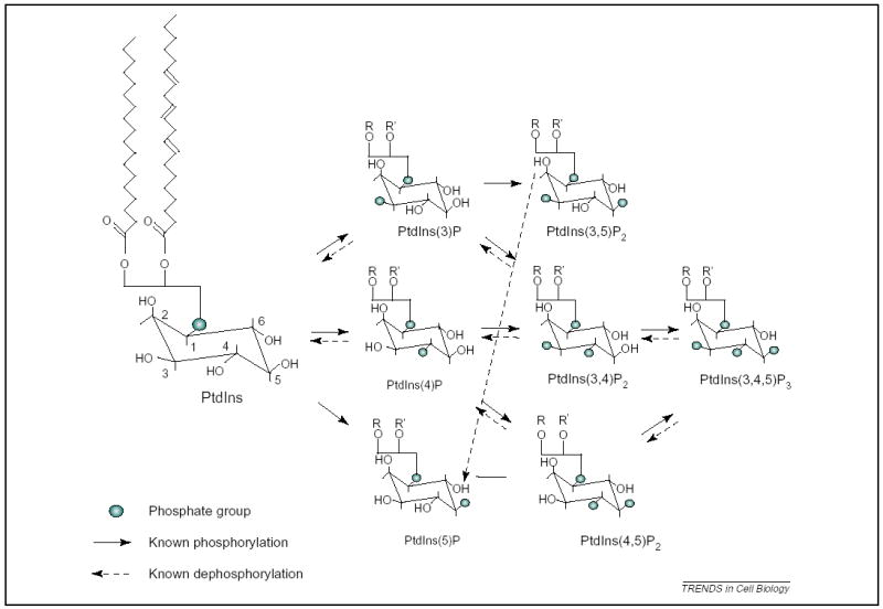
Phosphoinositide species. Phosphatidylinositol (PtdIns) consists of a D-myo-inositol 1-phosphate headgroup attached, almost exclusively, to 1-stearoyl, 2-arachanonyl, 3-phosphoglycerol as depicted. A variety of phosphoinositide kinases and phosphatases act to generate various phosphorylated derivatives, as indicated.
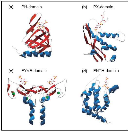
Phosphoinositide-binding domains. Structural examples of modular phosphoinositide-binding domains in complex with phosphoinositide-headgroup ligands and oriented such that the membrane interaction surface is top-most. Alpha-helices rendered in blue, beta-sheets in red. (a) Grp1 PH domain in complex with Ins(1,3,4,5)P4 [70]. (b) p40phox PX domain in complex with di-C4-PtdIns(3)P [71]. (c) Homodimeric EEA1 FYVE-domain (truncated to residues 1335–1411) in complex with Ins(1,3) P2. Green spheres represent Zn2+ ions [72]. (d) Epsin-1 ENTH-domain in complex with Ins(1,4,5)P3 [73]. Structures were manipulated in Deepview 3.7 and rendered using POV-ray 3.6.
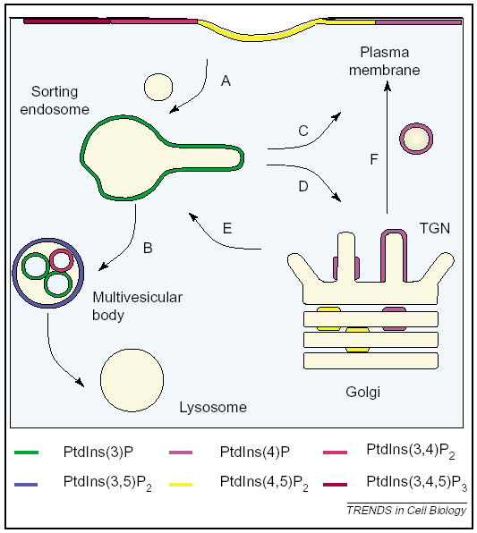
Cartoon depicting intracellular membranes in the endocytic and biosynthetic pathways and their hypothesized phosphoinositide content. These organelles control the trafficking of cargo to a variety of subcellular localizations. Organelle names given in type, trafficking pathways given by letters. A, internalization from the plasma membrane; B, degradative sorting to the lysosome; C, recycling from endosomes to the plasma membrane; D, retrieval of cargo from endosomes back to the trans-Golgi network (TGN); E, delivery of cargo from the TGN to endosomes; F, secretion of cargo from the TGN to the plasma membrane.
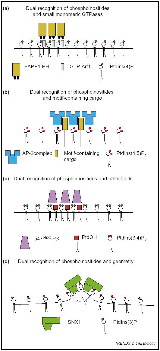
Coincidence detection to restrict localization. It is predicted that the following mechanisms will contribute towards the restricted localization of phosphoinositide-binding proteins across membrane. (a) Detection of phosphoinositides and small monomeric GTPases. Exemplified by the Arf1 and PtdIns(4)P-dependent localization of FAPP1 to the TGN. (b) Detection of phosphoinositides and cargo. Exemplified by the cargo and PtdIns(4,5)P2-dependent localization of the adaptor protein AP-2 complex to the plasma membrane. (c) Detection of phosphoinositides and other membrane lipids. Exemplified by the hypothetical PtdIns(3,4)P2- and PtdOH-dependent localization of p47phox. (d) Detection of phosphoinositides and geometric cues. Exemplified by the 3′-phosphoinositide and curvature-dependent localization of SNX1 to endosomes.
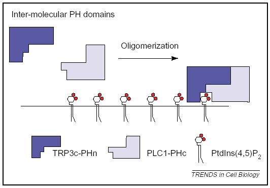
Intermolecular coincidence detection. Oligomerization of PLCγ1 and the TrpC3 Ca2+ channel creates a PtdIns(4,5)P2-binding PH domain from two half-PH domains.
References
-
- Cullen PJ, et al. Modular phosphoinositide-binding domains –their role in signalling and membrane trafficking. Curr Biol. 2001;11:R882–R893. - PubMed
-
- Lemmon MA. Phosphoinositide recognition domains. Traffic. 2003;4:201–213. - PubMed
-
- Balla T. Inositol-lipid binding motifs: signal integrators through protein–lipid and protein–protein interactions. J Cell Sci. 2005;118:2093–2104. - PubMed
-
- Roth MG. Phosphoinositides in constitutive membrane traffic. Physiol Rev. 2004;84:699–730. - PubMed
-
- Kobayashi T, et al. A lipid associated with the antiphospholipid syndrome regulates endosome structure and function. Nature. 1998;392:193–197. - PubMed
Publication types
MeSH terms
Substances
Grants and funding
LinkOut - more resources
Full Text Sources
