. 2005:64:231-61.
doi: 10.1016/S0065-3527(05)64007-9.

Mechanism of membrane fusion by viral envelope proteins

Affiliations

Mechanism of membrane fusion by viral envelope proteins

Stephen C Harrison. Adv Virus Res. 2005.

Abstract

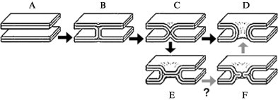
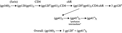
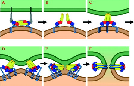
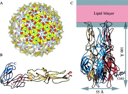
Enveloped viruses enter cells by fusing their lipid bilayer membrane with a cellular membrane. Most viral fusion proteins require priming by proteolytic processing, either of the fusion protein itself or of an accompanying protein. The priming step, which often occurs during transport of the fusion protein to the cell surface but may also occur extracellularly, then prepares the fusion protein for triggering by events that accompany attachment and uptake. Two classes of viral fusion proteins have been identified so far by structural studies. The fusion of two bilayers that these proteins catalyze is likely to proceed by the same pathway in both cases. That is, these proteins are like enzymes that have different structures but that still catalyze the same chemical reaction. It is found that bilayer fusion reaction is common to all the enveloped viral entry pathways. It is believed to pass through an intermediate known as a “hemifusion stalk.”


Figures

Fig 1
Fig 1
Fusion of two lipid bilayers. (A) Two parallel bilayer membranes. There is a substantial barrier to close approach. (B) Hemifusion stalk. (C) Proposed transition structure. (D) Fusion pore (before lateral expansion). (E) Hemifusion diaphragm. (F) Some models include perforation of the hemifusion diaphragm as a productive step toward fusion pore formation. Adapted from Jahn et al. (2003); see also Cohen et al. (2002).
Fig 2
Fig 2
The influenza virus hemagglutinin. (A) HA0, before cleavage between HA1 and HA2. The HA1 part of the protein is blue; the HA2 part, is purple. The fusion peptide, looped out before cleavage, is yellow. The sialic acid‐binding site and the cleavage point are shown by blue and yellow arrows, respectively. (B) The mature HA after cleavage but before low‐pH triggering. The only change from the structure in (A) is insertion of the fusion peptide (the N terminus of HA2, in yellow) into a crevice along the three‐fold axis (dark yellow asterisk). A purple arrow points to the loop between the shorter, N‐proximal helix and the longer, central helix. A purple asterisk indicates the position of residues that will move to the top of the molecule during the low pH‐induced transition. (C) HA2 after exposure to low pH. The same structure can be obtained by refolding HA2 expressed in E. coli. It is the minimal free‐energy state of HA2 unconstrained by covalent association with HA1. The long loop in the prefusion structure (purple arrow in [B]) has now become helical, elevating the N terminus of the protein (the fusion peptide itself is not included in this structure) to the top of the molecule (purple asterisk). A break and reversal of direction in the central α‐helix of the prefusion trimer likewise projects the C terminus of the protein to the top. The figure is aligned with respect to (B) so this break is roughly at the same height in both panels. In the actual transition, the chain reversal is likely to occur by melting and rezipping of the C‐terminal helical segment, as shown in Fig. 3C. For detailed references, see Skehel and Wiley (2000).
Fig 3
Fig 3
Diagram of membrane fusion mediated by class I viral fusion proteins. (A) Receptor binding (shown here for HIV or SIV Env, where the schematic receptor and coreceptor symbolize CD4 and CXCR4 or CCR5). (B) Dissociation of the receptor‐binding domain (in the case of HA, a disulfide bond prevents complete dissociation, but the structural rearrangement requires that HA1 move away from the threefold axis) and projection of the fusion peptide toward the target cell membrane. This state is known as the “prefusion intermediate” (or, sometimes, the “prehairpin intermediate”). The arrows show the positions along the core helical bundle at which, on HIV‐1 gp41, the inhibitor peptides T‐20 and C34 are expected to bind. (C) Folding back of the C‐terminal part of the molecule. This zipping up of a segment of the fusion protein sometimes designated “helical region 2” (HR2); helical region 1 (HR1) forms the inner core of the postfusion trimer) draws the two membranes together as the N‐terminal fusion peptide, inserted into the target cell membrane, and the C‐terminal transmembrane segment, which is anchored in the viral membrane, are forced by the refolding to approach each other. (D) Hemifusion stalk formation. Provided that they insert only into the outer leaflet of the bilayer, the fusion peptides can migrate into the stalk, as proposed here, and stabilize it. (E) Fusion pore formation. Hemifusion structures flicker transiently into unstable pores, which reseal. The pore can be trapped by the final refolding step, in which the three transmembrane segments snap into place around the inserted fusion peptides. In the case of HA, this step may be driven by formation of a set of interactions that cap the inner core helices.
Fig 4
Fig 4
Top: The ectodomain of influenza virus C HEF and its structural and apparent evolutionary relationship to influenza A HA. Bottom: A key to the color scheme for the HEF polypeptide chain. The N‐ and C‐ terminal segments of HEF1 and all of HEF2 (except for the fusion peptide) are in red. The acetylase enzymatic domain (E1 + E″ + E2) is in green; the receptor‐binding domain (R) is in blue. Think of the red fragments as an elementary fusion protein and the green and blue fragments as insertions. In HA, most of the acetylase domain has been deleted, except for the E′ fragment, which becomes an adaptor to connect the elementary fusion protein with the receptor‐binding domain. Adapted from Rosenthal et al. (1998).
Fig 5
Fig 5
States of the HIV‐1 envelope protein, as detected by biochemical, immunological, physicochemical, and structural analyses. The trimeric gp160 precursor is cleaved by a furin‐like protease to gp120 and gp41, which retain their threefold association. Binding of CD4 (one per trimer may be sufficient, but the degree of cooperativity is not determined) induces a conformational change in gp120 (gp120); binding of a suitable chemokine receptor (chR) may induce or lock in a further change (gp120). Ultimately gp120 is probably shed from the gp41 stem (gp120*). Once liberated, the gp41 trimer undergoes a two‐stage, fusion‐inducing conformational change: a transition to an extended, prefusion intermediate, (gp41)3, with the fusion peptide inserted into the target cell membrane, followed by a folding back to form the final, trimer‐of‐hairpins structure (gp41*)3, generating membrane fusion in the process.
Fig 6
Fig 6
Primary structure of the HIV and SIV envelope proteins. The bar labeled “gp160” represents, schematically, the regions of the envelope precursor. The parts of gp160 corresponding to gp120, gp41, and gp140 (the gp160 ectodomain) are shown as open bars. The scissors symbol shows the furin cleavage point. The gp120 core can be considered a receptor‐binding insertion into an elementary fusion protein, as diagrammed in the top two bars. Compare with Fig. 4—the gp120 core is the analog of the R and E′ regions of influenza HA. The principal elements of the gp41 ectodomain are diagrammed in the bottom bar.
Fig 7
Fig 7
Structure of the gp120 core in the CD4‐bound state (Kwong et al., 1998). (A) Ribbon representation, showing the inner and outer domains, linked by a bridging sheet. The locations of the V1–V2 and V3 loops, deleted from the core construct (compare with Fig. 6), are shown. The locations of carbohydrate chains are shown by molecular ball‐and‐stick representations of those sugars found to be well‐ordered in the crystal structure. The view is into the CD4‐binding pocket. N and C termini of the core are labeled. (B) Side view of the same structure, with the first two immunoglobulin‐like domains of CD4 also shown.
Fig 8
Fig 8
The inner core (HR1) and outer layer (HR2) of the gp41 ectodomain in the postfusion state of the gp41 trimer (Weissenhorn et al., 1997). The structure as determined crystallographically contains six peptides—three inner and three outer. The dashed lines show covalent connectivities that would be present in the intact gp41 trimer. The residue numbers (for gp41, counting from the N terminus of the fusion peptide) for the beginning and end of the inner and outer layer helices are shown for the red subunit.
Fig 9
Fig 9
The flavivirus fusion protein E. (A) Organization of E dimers in the virion surface. Each subunit is shown in three colors: domain I in red, domain II in yellow, and domain III in blue—based on the structure described by Kuhn et al., (2002). (B) The soluble ectodomain, sE of dengue virus type 2, in the dimeric prefusion conformation found on mature virions (Modis et al., 2003). The domain colors are as in (A). The bar above the ribbon diagram shows the relationship of domains to primary structure. The “stem” segment between residue 394 and the transmembrane anchor is not included in the three‐dimensional structure. (C) The sE trimer (Modis et al., 2004). The proteins are shown in relation to a schematic lipid bilayer to illustrate the likely degree of penetration of the fusion loops (top) into the membrane. The ribbon diagram (left) is colored as in (A) and (B). Arrows at the C terminus of the polypeptide chain suggest its presumed continuing direction. The surface rendering (right) includes a dashed arrow to show the proposed course of the stem peptide, which would lead to the transmembrane anchor.
Fig 10
Fig 10
Diagram of membrane fusion by class II viral fusion proteins. (A) Receptor binding through domain III of E (flaviviruses). (B) Lowered pH in an endosome leads to dissociation of the dimer interactions. On release of dimer constraints, monomers can flex outward, presenting their fusion loops to the target cell membrane. (C) Insertion of the fusion loops into the target cell membrane and initial formation of trimer contacts among the projecting domains II. (D) Domain III flips over and the stem zips up along the outside of the trimer. (E) Hemifusion stalk. The diagram shows a proposed role for the inserted fusion loops—stabilization of the hemifusion stalk. (F) Formation of a fusion pore. Completing the zipping up of the stem drives fusion forward, because the cytosolic tails enter the pore and commit it to dilation.
Fig 11
Fig 11
The alphavirus fusion protein E1. (A) Organization of E1 and E2 on the surface of virions. Simplified representations of subunits have been superimposed on a model of the fit of the SFV E1 crystal structure into an image reconstruction of the virion from electron cryomicroscopy (Lescar et al., 2001). E1 is red (domain I), yellow (domain II), and blue (domain III). The E2 trimer (for which only a 9‐Å structure is currently known, from electron microscopy) is represented by a green trefoil. It projects outward, capping the fusion loop of E1. Numbers (5, 3, and 2) show the positions of fivefold, threefold, and twofold icosahedral symmetry axes; triangles show the positions of local threefold axes in the T = 4 icosahedral surface lattice. (B) The soluble ectodomain, E1*, of Semliki Forest virus. Domain colors as in (A). (C) The E1* postfusion trimer. Each subunit is a single color. The stem of E1, shorter than the stem of flavivirus E proteins (compare with Fig. 9), would link the C terminus of E1* to the transmembrane anchor. (B) and (C) are adapted from Gibbons et al. (2004b).

Similar articles

Cited by

References

    1. Ahn A., Gibbons D.L., Kielian M. The fusion peptide of Semliki Forest virus associates with sterol‐rich membrane domains. J. Virol. 2002;76:3267–3275. - PMC - PubMed
    1. Allan J.S., Coligan J.E., Barin F., McLane M.F., Sodroski J.G., Rosen C.A., Haseltine W.A., Lee T.H., Essex M. Major glycoprotein antigens that induce antibodies in AIDS patients are encoded by HTLV‐III. Science. 1985;228:1091–1094. - PubMed
    1. Allison S.L., Schalich J., Stiasny K., Mandl C.W., Heinz F.X. Mutational evidence for an internal fusion peptide in flavivirus envelope protein E. J. Virol. 2001;75:4268–4275. - PMC - PubMed
    1. Allison S.L., Schalich J., Stiasny K., Mandl C.W., Kunz C., Heinz F.X. Oligomeric rearrangement of tick‐borne encephalitis virus envelope proteins induced by an acidic pH. J. Virol. 1995;69:695–700. - PMC - PubMed
    1. Allison S.L., Stiasny K., Stadler K., Mandl C.W., Heinz F.X. Mapping of functional elements in the stem‐anchor region of tick‐borne encephalitis virus envelope protein E. J. Virol. 1999;73:5605–5612. - PMC - PubMed

MeSH terms

LinkOut - more resources