Clinical Trial
. 2006 Jan 15;29(2):452-66.
doi: 10.1016/j.neuroimage.2005.07.048. Epub 2005 Sep 6.

Cytology and functionally correlated circuits of human posterior cingulate areas

Affiliations
Clinical Trial

Cytology and functionally correlated circuits of human posterior cingulate areas

Brent A Vogt et al. Neuroimage. .

Abstract

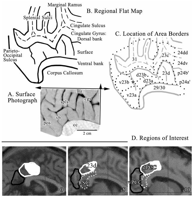
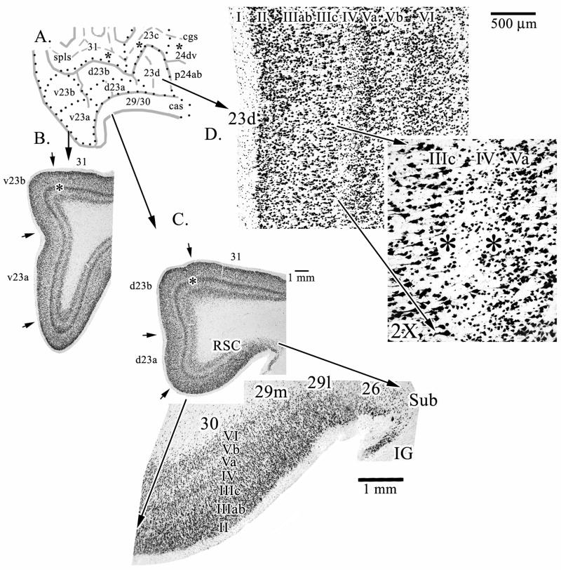
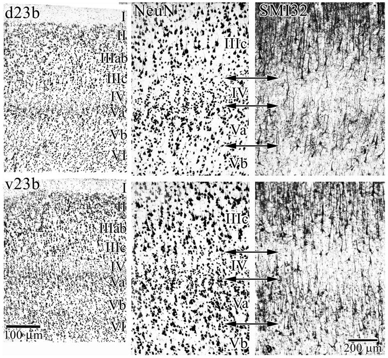
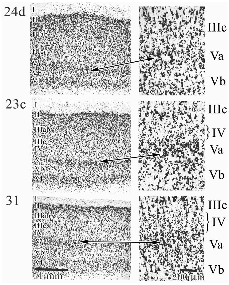
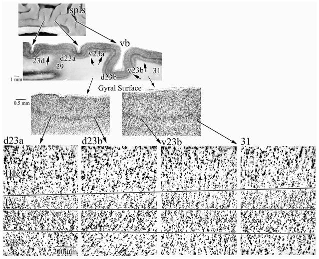
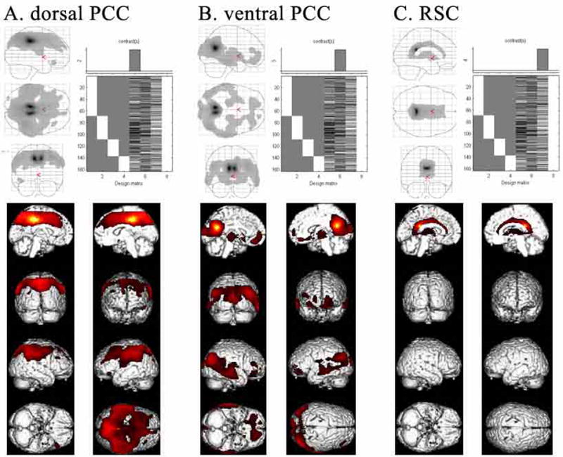
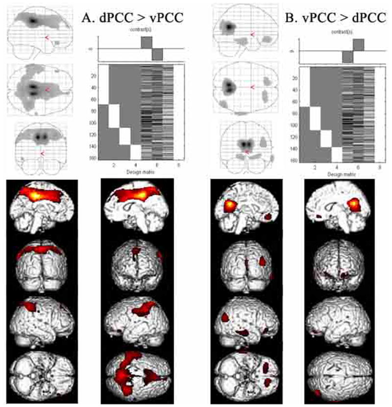
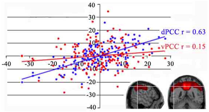
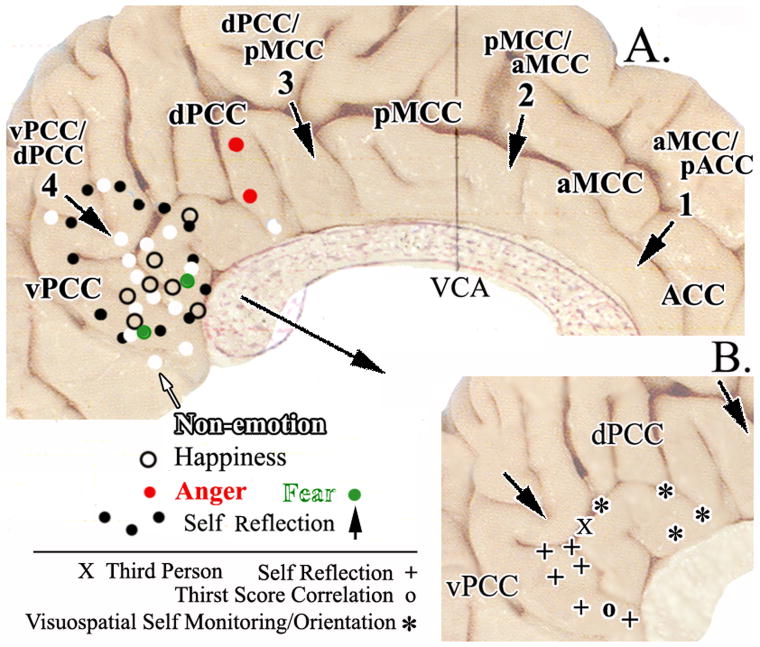
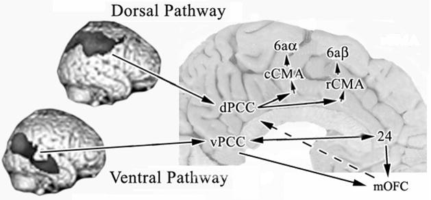
Human posterior cingulate cortex (PCC) and retrosplenial cortex (RSC) form the posterior cingulate gyrus, however, monkey connection and human imaging studies suggest that PCC area 23 is not uniform and atlases mislocate RSC. We histologically assessed these regions in 6 postmortem cases, plotted a flat map, and characterized differences in dorsal (d) and ventral (v) area 23. Subsequently, functional connectivity of histologically guided regions of interest (ROI) were assessed in 163 [(18)F]fluorodeoxyglucose human cases with PET. Compared to area d23, area v23 had a higher density and larger pyramids in layers II, IIIc, and Vb and more intermediate neurofilament-expressing neurons in layer Va. Coregisrtration of each case to standard coordinates showed that the ventral branch of the splenial sulci coincided with the border between d/v PCC at -5.4 +/- 0.17 cm from the vertical plane and +1.97 +/- 0.08 cm from the bi-commissural line. Correlation analysis of glucose metabolism using histologically guided ROIs suggested important circuit differences including dorsal and ventral visual stream inputs, interactions between the vPCC and subgenual cingulate cortex, and preferential relations between dPCC and the cingulate motor region. The RSC, in contrast, had restricted correlated activity with pericallosal cortex and thalamus. Visual information may be processed with an orbitofrontal link for synthesis of signals to drive premotor activity through dPCC. Review of the literature in terms of a PCC duality suggests that interactions of dPCC, including area 23d, orient the body in space via the cingulate motor areas, while vPCC interacts with subgenual cortex to process self-relevant emotional and non-emotional information and objects and self-reflection.


Figures

Figure 1
Figure 1
A. Medial surface photograph with planes of flattening in the dorsal (single arrow) and rostrocaudal (double arrow) orientations. B. Map of flattened sulci and gyri with the dorsal and ventral banks of the cingulate gyrus noted. The fundi of the cingulate and splenial sulci are dashed lines. The retrosplenial areas 29 and 30 were reflected ventrally below the edge of the corpus callosum so as to not interfere with showing area 23. C. Distribution of areas from a histological assessment (dotted lines) including an asterisk over the ventral branch (vb) of the splenial sulci (spls) where the border between areas d23b and v23b was measured for co-registration to standardized maps; also border #4 in Figure 6. The border between dPCC and vPCC is aligned with the vb and centered on the asterisk. D. Regions of Interest outlined in sagittal sections for the MNI brain (coordinates are the distance in mm from the midline) including dPCC (white outlines), area 23d (part of dPCC formed by solid white area inside the white outline), vPCC (black outlines), and retrosplenial cortex (RSC; white dots). cc, corpus callosum; cgs, cingulate sulcus; mr, marginal ramus; pos, parieto-occipital sulcus; spls, splenial sulci; vb, vertical branch of spls.
Figure 2
Figure 2
Overview of key components of the PCC and RSC regions. The three asterisks in the cgs and spls on the line drawing (A.) are where microphotographs were taken for sulcal areas in Figure 4. B. and C. are macrophotographs from levels through v23 and d23, respectively (asterisks emphasize layer Va at this magnification). A pull-out from C. further magnifies the retrosplenial areas 29 and 30 on the ventral bank of the cingulate sulcus. Area 26 is ectosplenial cortex. D. Dysgranular area 23d is shown with a pull-out magnification of a point at which the variability of layer IV (i.e., dysgranularity) is most evident. Between the asterisks it can be seen that layer IV discontinues for a short distance. Sub, dorsal subiculum; IG, indusium griseum.
Figure 3
Figure 3
Cytological details of the dorsal and ventral divisions of area 23b. The breadth and neuron densities in layers II, III, IV, and V are prominent. The density of NFP bearing neurons in layers IIIc, Va, and Vb are greater in v23b than in d23d as shown in the right panels (SMI32). Thus, phenotypic expression of NFP supports the dichotomy of PCC architecture observed in NeuN preparations.
Figure 4
Figure 4
Cingulate and splenial sulcal areas. Area 24d is provided as a comparison because it is agranular; i.e., lacks a layer IV and layers IIIc and Va directly abut each other. The arrow pair shows exactly which cortex was magnified at the right of the figure. In area 23c layers II–IV are quite broad and layer IV is present though thin. Area 31 has a well developed layer IV and the relative size of layer IIIc is greater than layer Va in contrast to other cingulate areas where there is a relative size equivalency among neurons in these layers. The brackets on layer IV emphasize difference in thickness in all areas.
Figure 5
Figure 5
Horizontal sections show the progressive differentiation of PCC in the rostrocaudal direction. The prominent ventral branch of the spls (vb) is shown in this section and areas d23a, d23b, v23b, and 31 are present. Notice that retrosplenial area 29 is cut with a glancing section at this level and no cytoarchitecture can be differentiated and the level of area 23a is quite dorsal and starting to merge with area 23b and the thickness of layer Va is increased. The highest magnification photographs were aligned on the layer IV/Va border and the other two borders emphasize the striking elaboration of layers IV and Va. Although each panel is a separate photograph, arrows are oriented to mark layer borders so the progressive differentiation of different layers can be compared and appreciated. For example, follow the progressive increase in width and neuron density of layer IV from the left to right of the figure.
Figure 6
Figure 6
ROIs based on histological assessment and co-registration (Fig. 1D) were used to evaluate CMRGlu in the resting state for 163 cases. SPM results were considered significant and are shown at false discovery rate (FDR)-corrected p value <0.001 for A. dPCC, B. vPCC, and C. RSC. The identified correlated voxels for each ROI suggest very different functional connectivity for each ROI.
Figure 7
Figure 7
Validation of the correlation analysis was performed by directly comparing bCCOI for each division of PCC to determine the level of significant difference between the two bCCOI. There is a reduction of medial prefrontal activity in dPCC and of posterior parietal activity in vPCC suggesting some functional overlap in these two regions. The thalamic site in vPCC is lost following interaction analysis with dPCC suggesting, that although thalamic activity was not observed in the dPCC correlations (Fig. 6), some may be present at a subthreshold level.
Figure 8
Figure 8
Plot of the regression between rCMRGlu in parietal cortex (x, y, z coordinates; −54, −44, 44 mm). Location of the parietal voxel is shown for two planes of section at the white cross hairs. A significant correlation is shown with dPCC but not for vPCC. Each dot represents one subject.
Figure 9
Figure 9
The four cingulate regions and their borders are shown with arrows and bold print in A (VCA, vertical plane at the anterior commissure). Activations associated with three simple emotions (happiness, anger, fear) generated with scripts or faces are shown as are those associated with non-emotional scripts and faces with symbols for peak activation sites discussed in the text. Overlap of emotional and non-emotional information processing suggests that vPCC has a nonspecific role in emotion that is not the case for subgenual ACC. A. also shows differentiation within PCC with the full area of activation in a study of self-reflection (black dots; Johnson et al., 2002). B. Pull out of the posterior cingulate region with locations of peak-voxel activations during self-reflection, third-person vs first person, and visuospatial self monitoring and orientation.
Figure 10
Figure 10
Differential information processing through the dorsal and ventral visual pathways to PCC based on presented human bCCOI and previous monkey corticocortical connection studies. A key function of PCC is in visuospatial orientation and visual processing pathways are preserved into PCC according to the bCCOI analysis. A circuit hypothesis of the flow of information through the cingulate gyrus is suggested to the rostral and caudal cingulate motor areas (rCMA, cCMA) to account for differences in functional activity for the two divisions of PCC and a terminal synthesis of information from the vPCC and ACC to medial orbitofrontal cortex (mOFC). A link to premotor systems (dashed arrow) is suggested by bCCOI in OFC with the dPCC ROI.

References

    1. Ashburner J, Andersson JL, Friston KJ. High-dimentional image registration using symmetric priors. NeuroImage. 1999;9:619–628. - PubMed
    1. Bentley DE, Derbyshire SWG, Youell PD, Jones AKP. Caudal cingulate cortex involvement in pain processing: an inter-individual laser evoked potential source localization study using realistic head models. Pain. 2003;102:265–271. - PubMed
    1. Bernstein LJ, Beig S, Siegenthaler AL, Grady CL. The effect of encoding strategy on the neural correlates of memory for faces. Neuropsychologia. 2002;40:86–89. - PubMed
    1. Berthoz A. Parietal and hippocampal contribution to topokinetic and topographic memory. Phil Trans Royal Soc Lond Series B: Biolog Sci. 1997;352:1437–1448. - PMC - PubMed
    1. Berthoz A. Hippocampal and parietal contribution to topokinetic and topographic memory. In: Burgess N, Jeffery KJ, O’Keefe J, editors. The Hippocampal and Parietal Foundations of Spatial Cognition. Oxford University Press; Oxford: 1999.

Publication types

MeSH terms