. 2005 Sep;79(18):11598-606.
doi: 10.1128/JVI.79.18.11598-11606.2005.

Antimicrobial peptides from amphibian skin potently inhibit human immunodeficiency virus infection and transfer of virus from dendritic cells to T cells

Affiliations

Antimicrobial peptides from amphibian skin potently inhibit human immunodeficiency virus infection and transfer of virus from dendritic cells to T cells

Scott E VanCompernolle et al. J Virol. 2005 Sep.

Abstract

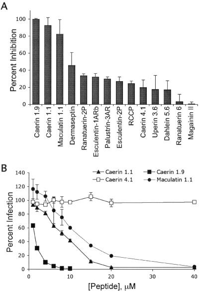
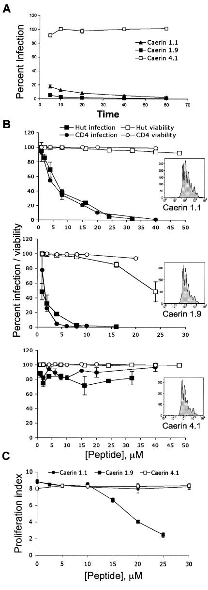
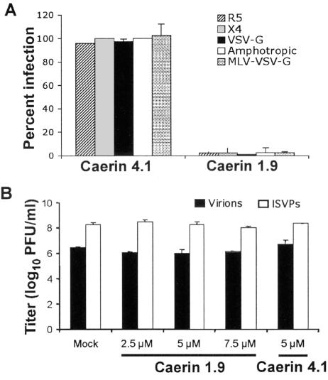
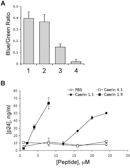
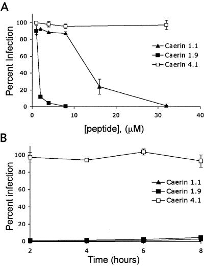
Topical antimicrobicides hold great promise in reducing human immunodeficiency virus (HIV) transmission. Amphibian skin provides a rich source of broad-spectrum antimicrobial peptides including some that have antiviral activity. We tested 14 peptides derived from diverse amphibian species for the capacity to inhibit HIV infection. Three peptides (caerin 1.1, caerin 1.9, and maculatin 1.1) completely inhibited HIV infection of T cells within minutes of exposure to virus at concentrations that were not toxic to target cells. These peptides also suppressed infection by murine leukemia virus but not by reovirus, a structurally unrelated nonenveloped virus. Preincubation with peptides prevented viral fusion to target cells and disrupted the HIV envelope. Remarkably, these amphibian peptides also were highly effective in inhibiting the transfer of HIV by dendritic cells (DCs) to T cells, even when DCs were transiently exposed to peptides 8 h after virus capture. These data suggest that amphibian-derived peptides can access DC-sequestered HIV and destroy the virus before it can be transferred to T cells. Thus, amphibian-derived antimicrobial peptides show promise as topical inhibitors of mucosal HIV transmission and provide novel tools to understand the complex biology of HIV capture by DCs.


Figures

FIG. 1.
FIG. 1.
Amphibian peptides inhibit HIV infection of T cells. (A) Fourteen purified peptides and one natural peptide mixture (RCCP) derived from the skin of anuran amphibians were incubated at 10 μg/ml (RCCP), 1 μM (ranatuerin-2P, esculentin-1ARb, palustrin-2AR, and esculentin-2P), 5 μM (caerin 1.9 and magainin II) or 10 μM (all others) with 3 × 10⁴ T cells and HIV-R5 at an MOI of 1 per cell at 37°C for 3 days. Cells were harvested and analyzed for GFP expression. The data are presented as the mean of three replicate samples from one representative experiment of two independent experiments. Error bars indicate standard deviations. Data are presented as percent inhibition normalized to infection in the presence of PBS. (B) T cells (3 × 10⁴) were incubated with HIV-R5 at an MOI of 1 and increasing concentrations of caerin 1.1, caerin 1.9, maculatin 1.1, caerin 4.1, or PBS at 37°C for 3 days. Cells were harvested and analyzed for GFP expression. The data are presented as the mean of three replicate samples from one representative experiment of six independent experiments. Error bars indicate standard deviations. IC50s were calculated by regression analysis.
FIG.2.
FIG.2.
Peptide inhibition occurs rapidly with no effect on cell viability. (A) Caerin 1.1 (20 μM), caerin 1.9 (5 μM), caerin 4.1 (20 μM), or PBS was incubated with 3 × 10⁴ IFU of HIV-R5. At the times shown, virus-peptide solutions were diluted to a concentration that resulted in minimal activity based the results shown in Fig. 1B (fourfold) in complete RPMI medium with 3 × 10⁴ T cells and incubated at 37°C for 3 days. Cells were harvested, fixed, and analyzed for GFP expression by flow cytometry. Data were normalized to infection following PBS treatment and are presented as the mean of three replicate samples from one representative experiment of three independent experiments, with error bars indicating standard deviation. (B) Peptides were incubated for 5 min at increasing concentrations (1 to 48 μM) with 5 × 10⁴ activated primary CD4+ T cells (circles) or Hut/CCR5 cells (squares) infected with HIV-R5 at an MOI of 1, diluted fourfold with complete RPMI medium, and incubated at 37°C for 3 days. Cells were harvested and analyzed for GFP expression (closed circles and squares). Data are normalized to infection following PBS treatment and are presented as the mean of three replicate samples from one representative experiment of three independent experiments with error bars indicating standard deviation. At 2 h postinfection, 2 × 10⁴ T cells were removed from the culture, stained with PI, and analyzed for viability by flow cytometry (open circles and squares). (C) Peptides were incubated for 5 min at increasing concentrations (1 to 50 μM) with 5 × 10⁴ CFSE-labeled primary CD4+ T cells. Cells were then diluted fourfold with complete RPMI medium and activated by T-cell receptor engagement as previously described (41). The activated cells were cultured for 4 days, and CFSE fluorescence intensity was measured by flow cytometry. Representative histograms (panel B, insets) from each peptide treatment are shown (40 μM caerin 1.1, 10 μM caerin 1.9, and 40 μM caerin 4.1), which were identical to those of the cells not treated with peptides.
FIG. 3.
FIG. 3.
Peptide activity against HIV with different envelopes and diverse viral species. (A) Caerin 1.9 (5 μM) and caerin 4.1 (5 μM) were incubated with 3 × 10⁴ T cells and HIV-R5, HIV-X4, HDV-VSV-G, or HDV pseudotyped with the MLV amphotropic envelope at an MOI of 1 at 37°C for 3 days. Caerin 1.9 (10 μM) and caerin 4.1 (10 μM) were added to MLV (30,000 IFU) and incubated at 37°C for 45 min. Virus was then added to 3 × 10⁴ Mus dunni cells and cultured at 37°C for 3 days. Following incubation, cells were harvested and analyzed for GFP expression by flow cytometry. Data are normalized to infection following PBS treatment and are presented as the mean of three replicate samples from one representative experiment of three independent experiments, with error bars indicating standard deviation. (B) Reovirus T1L virions or ISVPs were incubated with caerin 1.9, caerin 4.1, or PBS (mock) at the concentrations shown at 37°C for 45 min. Virus was adsorbed to confluent HeLa cell monolayers at an MOI of 2 PFU per cell at 4°C for 1 h. Following two washes with PBS, fresh medium was added, and cells were incubated at 37°C for 24 h. Cells and medium were subjected to three cycles of freezing and thawing, and virus titer was determined by plaque assay. The results are presented as the means of two independent experiments. Error bars represent standard deviations.
FIG. 4.
FIG. 4.
Caerin 1.1 and caerin 1.9 disrupt the HIV envelope. (A) HIV-R5 carrying a β-lactamase reporter protein was incubated with 5 μM caerin 1.9 (lane 3) or PBS (lane 1) for 1 h prior to inoculation of Hut/CCR5 cells (105) by centrifugation at 600 × g for 1.5 h at 35°C. These cells or uninoculated cells (lane 4) were incubated at 37°C for 2 h to allow virus-cell fusion. CCF2/AM (20 μM) was added, the cultures were incubated at room temperature for 14 h, and fluorescence was measured at 447 and 520 nm by using a microplate fluorometer after excitation at 409 nm. Fluorescence ratios were calculated after subtraction of the average background fluorescence and are presented as relative fusion. Caerin 1.9 (5 μM) was added during the CCF2/AM incubation to control for peptide effects on dye loading (lane 2). Duplicate determinations were performed for each virus dilution. (B) HIV-VSV-G was incubated with peptides or PBS at the concentrations shown for 30 min in complete RPMI. Medium was then diluted 1:1,000 in assay buffer without detergent and analyzed for the presence of viral core protein p24 by ELISA. Total p24 was calculated from dilutions of replicate samples using linear regression analysis from p24 standards included on each plate. Data are presented as the mean of two replicate samples and are representative of two experiments. Error bars indicate standard deviation.
FIG. 5.
FIG. 5.
Amphibian antimicrobial peptides inhibit DC-mediated trans infection of T cells. (A) DCs (4 × 10⁴) were pulsed with HIV-R5 at an MOI of 2. Virus-cell mixtures were centrifuged for 1 h at 2,000 rpm and incubated at 37°C for 2 h to allow DCs to efficiently capture the virus. DCs were washed three times with complete RPMI medium to remove unbound virus. Peptide was then added to the DCs at the concentrations shown and incubated at 37°C for 45 min. DCs were washed and incubated with 3 × 10⁴ T cells at 37°C for 3 days. Cells were harvested, fixed with 1% paraformaldehyde, and analyzed for GFP expression. Data are normalized to infection following PBS treatment and are presented as the mean of three replicate samples from one representative experiment of three independent experiments, with error bars indicating standard deviation. (B) DCs (4 × 10⁴) were incubated with HIV-R5 at an MOI of 1. At the times shown postinoculation, 30 μM caerin 1.1, 4 μM caerin 1.9, and 30 μM caerin 4.1 were added to cultures and incubated at 37°C for 45 min. DCs were washed three times in complete RPMI medium, incubated with 2 × 10⁴ T cells at 37°C for 3 days, and analyzed for GFP expression. Data are normalized as above and are representative of four independent experiments.

References

    1. Ali, M. F., K. R. Lips, F. C. Knoop, B. Fritzsch, C. Miller, and J. M. Conlon. 2002. Antimicrobial peptides and protease inhibitors in the skin secretions of the crawfish frog, Rana areolata. Biochim. Biophys. Acta 1601:55-63. - PubMed
    1. Baer, G. S., and T. S. Dermody. 1997. Mutations in reovirus outer-capsid protein σ3 selected during persistent infections of L cells confer resistance to protease inhibitor E64. J. Virol. 71:4921-4928. - PMC - PubMed
    1. Balla, M. S., J. H. Bowie, and F. Separovic. 2004. Solid-state NMR study of antimicrobial peptides from Australian frogs in phospholipid membranes. Eur. Biophys. J. 33:109-116. - PubMed
    1. Barton, E. S., J. C. Forrest, J. L. Connolly, J. D. Chappell, Y. Liu, F. Schnell, A. Nusrat, C. A. Parkos, and T. S. Dermody. 2001. Junction adhesion molecule is a receptor for reovirus. Cell 104:441-451. - PubMed
    1. Belaid, A., M. Aouni, R. Khelifa, A. Trabelsi, M. Jemmali, and K. Hani. 2002. In vitro antiviral activity of dermaseptins against herpes simplex virus type 1. J. Med. Virol. 66:229-234. - PubMed

Publication types

MeSH terms

Substances