. 2005 Sep;79(18):11705-15.
doi: 10.1128/JVI.79.18.11705-11715.2005.

Sialidase activity of influenza A virus in an endocytic pathway enhances viral replication

Affiliations

Sialidase activity of influenza A virus in an endocytic pathway enhances viral replication

Takashi Suzuki et al. J Virol. 2005 Sep.

Abstract

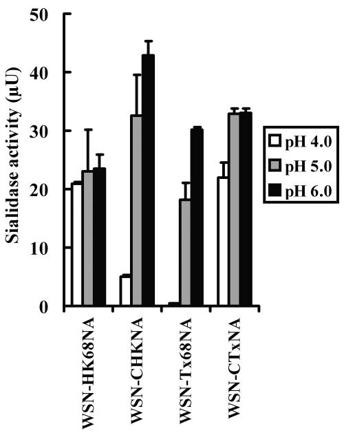
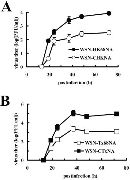
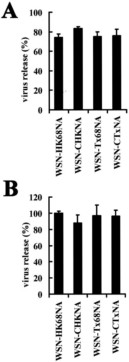
N2 neuraminidase (NA) genes of the 1957 and 1968 pandemic influenza virus strains possessed avian-like low-pH stability of sialidase activity, unlike most epidemic strains. We generated four reverse-genetics viruses from a genetic background of A/WSN/33 (H1N1) that included parental N2 NAs of 1968 pandemic (H3N2) and epidemic (H2N2) strains or their counterpart N2 NAs in which the low-pH stability of the sialidase activity was changed by substitutions of one or two amino acid residues. We found that the transfectant viruses bearing low-pH-stable sialidase (WSN/Stable-NAs) showed 25- to 80-times-greater ability to replicate in Madin-Darby canine kidney (MDCK) cells than did the transfectant viruses bearing low-pH-unstable sialidase (WSN/Unstable-NAs). Enzymatic activities of WSN/Stable-NAs were detected in endosomes of MDCK cells after 90 min of virus internalization by in situ fluorescent detection with 5-bromo-4-chloro-indole-3-yl-alpha-N-acetylneuraminic acid and Fast Red Violet LB. Inhibition of sialidase activity of WSN/Stable-NAs on the endocytic pathway by pretreatment with 4-guanidino-2,4-dideoxy-N-acetylneuraminic acid (zanamivir) resulted in a significant decrease in progeny viruses. In contrast, the enzymatic activities of WSN/Unstable-NAs, the replication of which had no effect on pretreatment with zanamivir, were undetectable in cells under the same conditions. Hemadsorption assays of transfectant-virus-infected cells revealed that the low-pH stability of the sialidase had no effect on the process of removal of sialic acid from hemagglutinin in the Golgi regions. Moreover, high titers of viruses were recovered from the lungs of mice infected with WSN/Stable-NAs on day 3 after intranasal inoculation, but WSN/Unstable-NAs were cleared from the lungs of the mice. These results indicate that sialidase activity in late endosome/lysosome traffic enhances influenza A virus replication.


Figures

FIG. 1.
FIG. 1.
Low-pH stability of sialidase activities of transfectant viruses generated by a reverse-genetics system. The amount of each transfectant virus (WSN-HK68NA, WSN-CHKNA, WSN-Tx68NA, and WSN-CTxNA) tested was determined by Western blotting techniques with anti-N2 NA monoclonal antibodies. The low-pH stability of sialidase activities of the viruses at pH 4.0, 5.0, or 6.0 was examined by using 4-MUα-Neu5Ac as described in Materials and Methods.
FIG. 2.
FIG. 2.
Growth kinetics of the transfectant viruses on MDCK cells. MDCK cells were infected with each transfectant virus at an MOI of 0.002 PFU/cell, and the culture media were harvested at indicated times after infection. The virus titers at each time point are the averages and the standard deviations of results of duplicate experiments. (A) Growth curves of the transfectant viruses, including parental N2 NA of pandemic A/Hong Kong/1/68 (H3N2) (•; WSN-HK68NA) or including its counterpart N2 NA (○; WSN-CHKNA); (B) growth curves of the transfectant viruses, including parental N2 NA of epidemic A/Texas/68 (H2N2) (▪); WSN-CTx68NA) or including its counterpart N2 NA (□; WSN-CTxNA).
FIG. 3.
FIG. 3.
Release of transfectant viruses from MDCK cells. MDCK cells were infected with each transfectant virus at an MOI of 1 PFU/cell. As controls, MDCK cells, which were infected with each transfectant virus under the same conditions, were treated with bacterial sialidases for 1 h at 37°C to release all budded viruses from the cell surface. Virus titers in each supernatant were determined by a plaque assay on MDCK cells. The amount of virus released at 13 h (A) or 24 h (B) postinfection was calculated as a percentage of virus titers with treatment of bacterial sialidases.
FIG. 4.
FIG. 4.
Plaque formation by the transfectant viruses on MDCK cells. MDCK cell monolayers were infected with each transfectant virus. Infected monolayers were overlaid with 2 ml of a solution of MEM containing acetylated trypsin (1 μg/ml) and 0.8% agarose. After incubation for 3 days at 34.5°C, plaques were fixed and stained with 0.5% amide black solution.
FIG. 5.
FIG. 5.
In situ fluorescent detection of sialidase activity of the transfectant virus NA in MDCK cells. (A) MDCK cells grown on glass coverslips were adsorbed with each transfectant virus at an MOI of 100 PFU/cell on ice for 90 min. After incubation at 37°C for 5 min or 90 min, the cells were fixed and permeabilized with cold methanol for 5 min. (B) MDCK cells were adsorbed with WSN-HK68NA or WSN-CTxNA in the same way. After incubation at 37°C for 90 min, the cells were fixed and incubated with or without 5 μM zanamivir in 0.1 M sodium acetate buffer (pH 5.0) for 5 min. The cells were observed with a laser scanning confocal microscope. Viral sialidase activities (red) were detected with X-Neu5Ac and Fast Red Violet LB as described in Materials and Methods. Early endosomes (green) were detected by a monoclonal antibody against EEA1 and FITC-conjugated goat anti-mouse IgG+M antibody. The nuclei (blue) were visualized by DAPI staining.
FIG. 6.
FIG. 6.
Comparison of the levels of adsorption of transfectant viruses to MDCK cells and their sialidase activities under the endocytosis pathway by FACS analysis. MDCK cells were adsorbed with each transfectant virus on ice for 90 min. The cells were fixed and permeabilized just after adsorption and after 90 min postinfection at 37°C as described in Materials and Methods. (A) The transfectant viruses adsorbed to the cells were detected with anti-H1N2 polyclonal antibody. (B) Viral sialidase activities in the cells at 90 min postinfection were detected with X-Neu5Ac and Fast Red Violet LB. Each experiment was performed twice; representative data are shown. The mean and median fluorescence intensities are shown in the panels. WSN-HK68NA, red; WSN-CHKNA, blue; WSN-Tx68NA, green; WSN-CTxNA, yellow; mock, black.
FIG. 7.
FIG. 7.
Suppression of infectious progeny virus by inhibition of sialidase activities of transfectant virus NAs under endocytosis. MDCK cells were treated with 5 μM zanamivir and 0.001% DEAE-dextran up to 3.5 h postinfection. After treatment with zanamivir, the cells were washed thoroughly to exclude residual zanamivir. The culture media (A) were harvested at 18 h after the infection of each transfectant virus, and the cells were fixed with cold methanol. As controls, MDCK cells were treated with zanamivir after 4 h of virus infection, and the media (D) were harvested at 18 h after infection. Progeny virus titers in the media (A and D) and the viral-antigen-positive cells (B) are shown as percentages of that of each counterpart control without zanamivir. Progeny virus titers per viral-antigen-positive cell (C) were calculated to estimate the effect of zanamivir on viral replication among the four transfectant viruses.
FIG. 8.
FIG. 8.
Effect of zanamivir on hemadsorption of MDCK and COS7 cells infected with the transfectant viruses. MDCK and COS7 cells were infected with each transfectant virus. After incubation with 5 μM zanamivir for 13 h, hemadsorption of the cells was determined by absorbance at 545 nm and is shown as a percentage relative to the hemadsorption of cells incubated without zanamivir. Each value is the mean from triplicate experiments.

References

    1. Clague, M. J. 1998. Molecular aspects of the endocytic pathway. Biochem. J. 336:271-282. - PMC - PubMed
    1. Garcia-Sastre, A., R. K. Durbin, H. Zheng, P. Palese, R. Gertner, D. E. Levy, and J. E. Durbin. 1998. The role of interferon in influenza virus tissue tropism. J. Virol. 72:8550-8558. - PMC - PubMed
    1. Goto, H., and Y. Kawaoka. 1998. A novel mechanism for the acquisition of virulence by a human influenza A virus. Proc. Natl. Acad. Sci. USA 95:10224-10228. - PMC - PubMed
    1. Hatta, M., P. Gao, P. Halfmann, and Y. Kawaoka. 2001. Molecular basis for high virulence of Hong Kong H5N1 influenza A viruses. Science 293:1840-1842. - PubMed
    1. Henkel, J. R., and O. A. Weisz. 1988. Influenza virus M2 protein slows traffic along the secretory pathway. pH perturbation of acidified compartments affects early Golgi transport steps. J. Biol. Chem. 273:6518-6524. - PubMed

Publication types

MeSH terms

LinkOut - more resources