. 2005 Sep;79(18):11742-51.
doi: 10.1128/JVI.79.18.11742-11751.2005.

Rab9 GTPase is required for replication of human immunodeficiency virus type 1, filoviruses, and measles virus

Affiliations

Rab9 GTPase is required for replication of human immunodeficiency virus type 1, filoviruses, and measles virus

James L Murray et al. J Virol. 2005 Sep.

Abstract

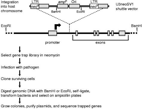
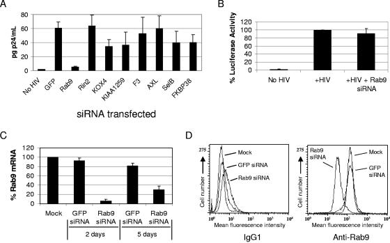
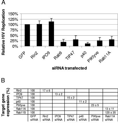
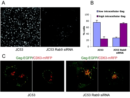
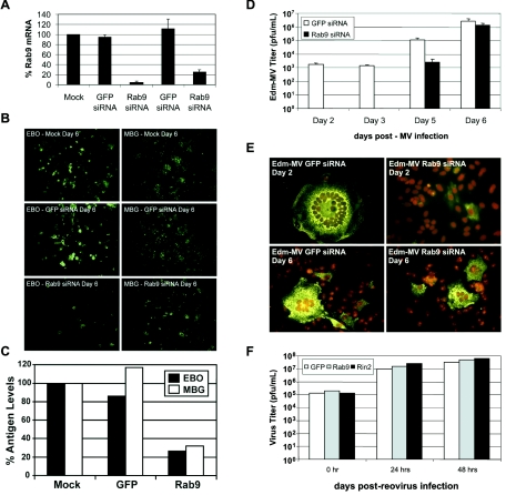
Rab proteins and their effectors facilitate vesicular transport by tethering donor vesicles to their respective target membranes. By using gene trap insertional mutagenesis, we identified Rab9, which mediates late-endosome-to-trans-Golgi-network trafficking, among several candidate host genes whose disruption allowed the survival of Marburg virus-infected cells, suggesting that Rab9 is utilized in Marburg replication. Although Rab9 has not been implicated in human immunodeficiency virus (HIV) replication, previous reports suggested that the late endosome is an initiation site for HIV assembly and that TIP47-dependent trafficking out of the late endosome to the trans-Golgi network facilitates the sorting of HIV Env into virions budding at the plasma membrane. We examined the role of Rab9 in the life cycles of HIV and several unrelated viruses, using small interfering RNA (siRNA) to silence Rab9 expression before viral infection. Silencing Rab9 expression dramatically inhibited HIV replication, as did silencing the host genes encoding TIP47, p40, and PIKfyve, which also facilitate late-endosome-to-trans-Golgi vesicular transport. In addition, silencing studies revealed that HIV replication was dependent on the expression of Rab11A, which mediates trans-Golgi-to-plasma-membrane transport, and that increased HIV Gag was sequestered in a CD63+ endocytic compartment in a cell line stably expressing Rab9 siRNA. Replication of the enveloped Ebola, Marburg, and measles viruses was inhibited with Rab9 siRNA, although the non-enveloped reovirus was insensitive to Rab9 silencing. These results suggest that Rab9 is an important cellular target for inhibiting diverse viruses and help to define a late-endosome-to-plasma-membrane vesicular transport pathway important in viral assembly.


Figures

FIG. 1.
FIG. 1.
Identification of cellular genes required for viral replication by gene trap mutagenesis. Susceptible host cell lines are chosen that are normally killed by the virus being studied. Cells are infected with the U3neoSV1 retrovirus (MOI = 0.1) carrying promoterless neomycin resistance genes (neor) in each LTR. Integration of the U3neoSV1 shuttle vector between an active promoter and an early exon results in gene disruption (trapping), with concomitant expression of the neor gene from the upstream (5′) LTR. The promoterless 3′ LTR is transcriptionally inactive. Following neomycin selection, the resultant gene trap library is infected with the virus of choice. Cell survival following viral infection results from trapping genes that are necessary for viral replication. Surviving cells are expanded and cloned. The shuttle vector is then rescued from genomic DNA as indicated above, and the recovered chromosomal DNA flanking the shuttle vector integration site is sequenced to identify the trapped gene.
FIG. 2.
FIG. 2.
Inhibition of HIV-1 replication with Rab9 siRNA. The JC53 cells used for HIV p24 assays are CD4+ CCR5+ CXCR4+ HeLa-derived indicator cells that express luciferase following HIV infection. Luciferase expression from a stably integrated HIV-LTR-luciferase construct is regulated by the early HIV gene product Tat. (A) JC53 HeLa cells were transfected with the indicated siRNAs and inoculated with HIV-1 LAV. HIV p24 secreted into culture supernatants was quantitated in p24 assays at 3 days postinoculation (n = 6). (B) Luciferase assays (n = 5) were performed to quantitate the infection of JC53 cells following Rab9 siRNA transfections. (C) Total mRNA isolated from JC53 cells (at 2 and 5 days posttransfection with GFP or Rab9 siRNA) was reverse transcribed, and the relative levels of Rab9 message originally present were determined by real-time PCR and normalized to HGPRT expression (n = 4). (D) Flow cytometric analysis of Rab9 protein expression following the indicated siRNA transfections. Staining was performed using a mouse anti-Rab9 antibody or a mouse IgG1 negative control.
FIG. 3.
FIG. 3.
HIV utilization of host genes facilitating vesicular transport. (A) JC53 cells were transfected with the indicated siRNAs for 48 h, inoculated with HIV (LAV strain; MOI = 1), and expanded in duplicate T75 flasks. Culture supernatants were harvested at 3 days postinoculation, and HIV p24 assays were performed. Values were normalized to HIV p24 secretion from GFP siRNA-transfected cells and represent the results of at least four independent experiments. (B) JC53 cells were transfected with siRNAs against the target genes specified, and the effect on target gene expression was determined as described in the legend to Fig. 2C.
FIG. 4.
FIG. 4.
Colocalization of CD63 and HIV Gag in Rab9-depleted cells. (A) Rab9 expression was visualized in parental JC53 cells (left panel) and JC53 Rab9 siRNA cells (right panel) by sequential incubations with a primary anti-Rab9 antibody and an AF488-conjugated secondary antibody. (B) JC53 cells and JC53 Rab9 siRNA cells were transfected with pGag-EGFP and pCD63-mRFP plasmids, and 20 to 60 cells per experiment (n = 3) were optically z-sectioned live at 36 h posttransfection. Cells having <10 or ≥10 intracellular Gag-EGFP puncta colocalizing with CD63-mRFP were classified as having low or high intracellular Gag, respectively. (C) Representative confocal sections of JC53 cells (left panels) and JC53 Rab9 siRNA cells (right panels) cotransfected with pGag-EGFP (green) and pCD63-mRFP (red).
FIG. 5.
FIG. 5.
Effects of Rab9 silencing on Ebola virus, Marburg virus, measles virus, and reovirus replication. All assays were performed in Vero cells. (A) Rab9 silencing in Vero cells. Rab9 mRNA expression in mock- or siRNA-transfected Vero cells was determined at 2 or 5 days posttransfection, as described in the legend to Fig. 2C (n = 3). (B) At 2 days posttransfection, mock- or siRNA-transfected Vero cells were inoculated with Ebola virus (EBO) (left panels) or Marburg virus (MBG) (right panels), and viral antigen production was visualized at 6 days postinoculation using polyclonal antisera against Ebola virus or Marburg virus antigens and a FITC-labeled secondary antibody. Magnification, ×20. (C) Antigen-capture assays. Quantitation of viral antigens released into culture supernatants (at 6 days postinoculation) was performed using an enzyme-linked immunosorbent assay-based antigen-capture assay (n = 2). (D) Viral titers of measles virus strain Edmonston (Edm-MV) released into the supernatants of GFP and Rab9 siRNA Vero cell transfectants (n = 3). (E) Measles virus Edmonston immunofluorescence staining. Measles virus syncytium formation was visualized by immunostaining of the measles virus nucleoprotein (green) at 2 and 6 days postinoculation. Cell nuclei were stained with propidium iodide (red). Magnification, ×200. (F) Reovirus assays. Vero cell GFP, Rab9, and Rin2 siRNA transfectants were inoculated for 1 h with reovirus type 1, washed, and grown for 0 to 2 days postinoculation before freezing at −20°C. Cell lysates were titrated by standard plaque assays on L-cell monolayers. Results from a single experiment are shown and are representative of three independent experiments.
FIG. 6.
FIG. 6.
Alternative pathways for HIV viral egress. HIV Env and Gag are expressed on the cell surface before internalization to the late endosome (LE), an important site of assembly initiation. (A) In macrophages, HIV buds into the lumen of the late endosome to form multivesicular bodies (MVBs), which can be released as exosomes by multivesicular body fusion with the plasma membrane. (B) In T cells and cultured cell lines, budding occurs primarily at the plasma membrane. Vesicular trafficking of viral proteins out of the late endosome to the trans-Golgi network (TGN) and plasma membrane may be aided, as indicated, by the host proteins Rab9, Tip47, p40, PIKfyve, and Rab11A. Rab11A may also favor plasma membrane budding by taking part in the recycling of viral proteins back to the plasma membrane via early endosomes (EEs) and recycling endosomes (REs).

Similar articles

Cited by

References

    1. Avota, E., N. Muller, M. Klett, and S. Schneider-Schaulies. 2004. Measles virus interacts with and alters signal transduction in T-cell lipid rafts. J. Virol. 78:9552-9559. - PMC - PubMed
    1. Basyuk, E., T. Galli, M. Mougel, J. M. Blanchard, M. Sitbon, and E. Bertrand. 2003. Retroviral genomic RNAs are transported to the plasma membrane by endosomal vesicles. Dev. Cell 5:161-174. - PubMed
    1. Bavari, S., C. M. Bosio, E. Wiegand, G. Ruthel, A. B. Will, T. W. Geisbert, M. Hevey, C. Schmaljohn, A. Schmaljohn, and M. J. Aman. 2002. Lipid raft microdomains: a gateway for compartmentalized trafficking of Ebola and Marburg viruses. J. Exp. Med. 195:593-602. - PMC - PubMed
    1. Becker, M. M., M. I. Goral, P. R. Hazelton, G. S. Baer, S. E. Rodgers, E. G. Brown, K. M. Coombs, and T. S. Dermody. 2001. Reovirus sigmaNS protein is required for nucleation of viral assembly complexes and formation of viral inclusions. J. Virol. 75:1459-1475. - PMC - PubMed
    1. Blot, G., K. Janvier, S. Le Panse, R. Benarous, and C. Berlioz-Torrent. 2003. Targeting of the human immunodeficiency virus type 1 envelope to the trans-Golgi network through binding to TIP47 is required for env incorporation into virions and infectivity. J. Virol. 77:6931-6945. - PMC - PubMed

Publication types

MeSH terms

LinkOut - more resources