. 2005 Sep 1;19(17):2078-90.
doi: 10.1101/gad.338505.

Identification of the scaramanga gene implicates Neuregulin3 in mammary gland specification

Affiliations

Identification of the scaramanga gene implicates Neuregulin3 in mammary gland specification

Beatrice Howard et al. Genes Dev. .

Abstract

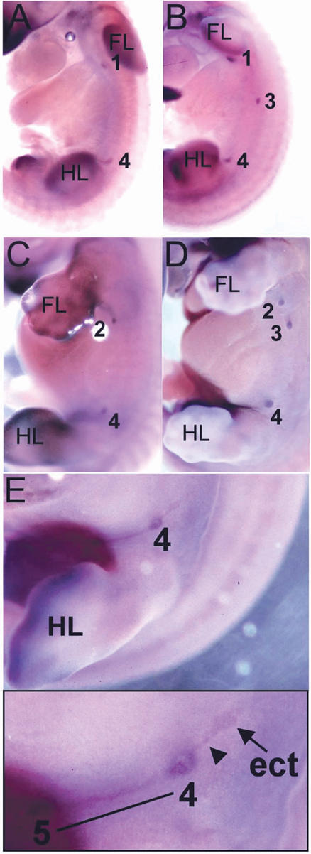
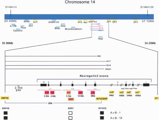
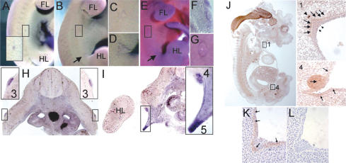
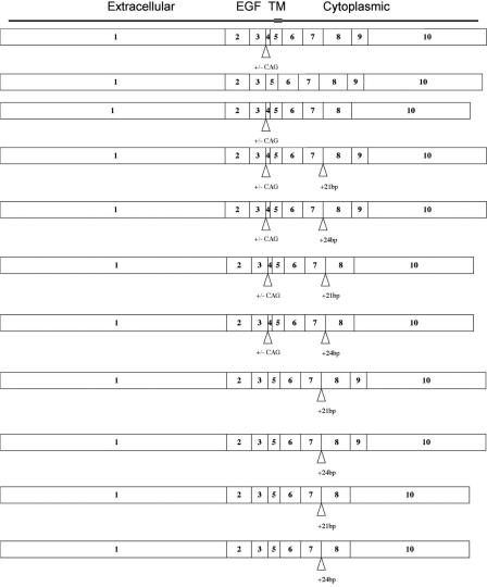
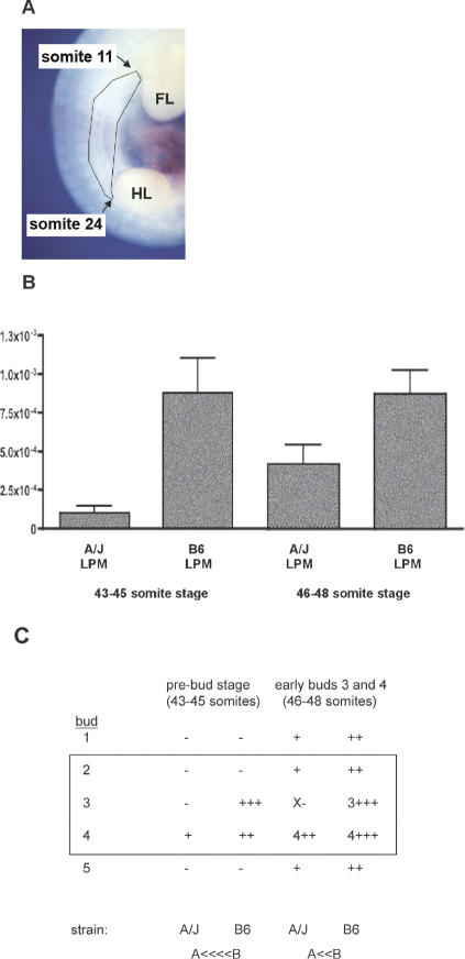
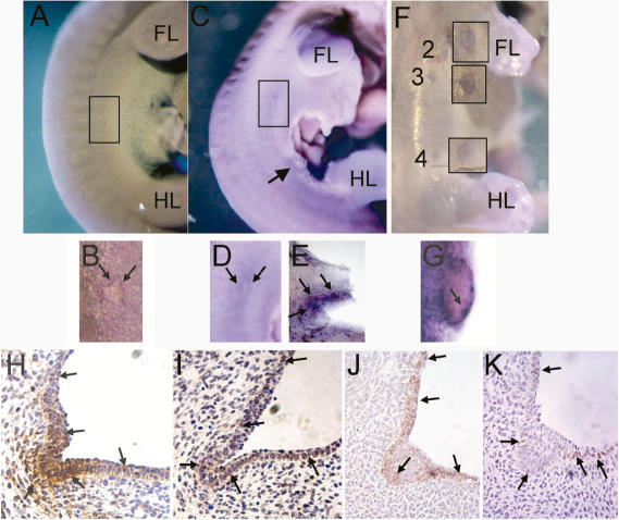
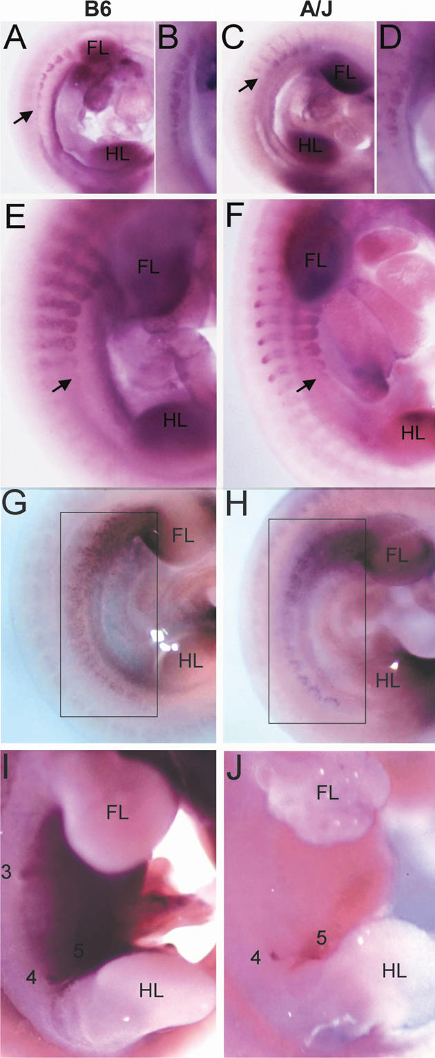
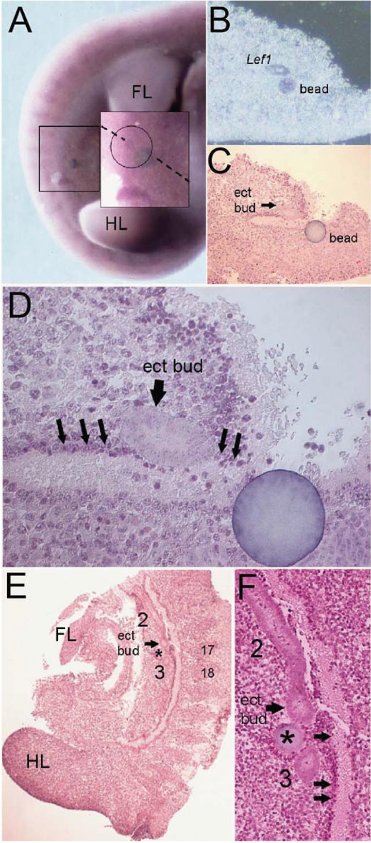
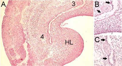
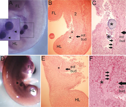
The mouse scaramanga (ska) mutation impairs mammary gland development such that both abrogation and stimulation of gland formation occurs. We used positional cloning to narrow the interval containing scaramanga (ska) to a 75.6-kb interval containing the distal part of the Neuregulin3 (Nrg3) gene. Within this region the only sequence difference between ska and wild-type mice is in a microsatellite repeat within intron 7. This alteration correlates with variations in Nrg3 expression profiles both at the whole embryo level and locally in the presumptive mammary region in ska mice. Localized expression of Nrg3 and its receptor, Erbb4, in the presumptive mammary region around the future bud site prior to morphological appearance of buds and the expression of bud epithelial markers further support an inductive role. Finally, Neuregulin3 (Nrg3)-soaked beads can induce expression of the early bud marker Lef1 in mouse embryo explant cultures, and epithelial bud formation can be observed histologically, suggesting that initiation of mammary bud development occurs. Taken together, these results indicate that a Neuregulin signaling pathway is involved in specification of mammary gland morphogenesis and support the long-held view that mesenchymal signal(s) are responsible for mammary gland inductive/initiating events.


Figures

Figure 1.
Figure 1.
Mammary bud 3 is absent and ectopic mammary buds form in A/J mice. Lef1 expression marks the position of mammary buds. Numbers denote the positions of the five mammary gland anlage. (FL) Forelimb bud; (HL) hindlimb bud. (A,B) Mammary buds are first observed in 48-somite embryos when bud 2 has not yet formed. Mammary bud 3 has not formed in an A/J embryo (A) shown by comparison with B6 (B). (C-E) Mammary buds in E12.5 embryos. (C) A 53-somite A/J embryo with absent bud 3. (D) A 55-somite B6 embryo. (E) A 54-somite A/J embryo with a presumptive supernumerary mammary bud adjacent to mammary bud 4. This is shown in magnified view in the inset. The ectopic Lef1-expressing cells are denoted by an arrow and a line of Lef1-expressing cells connecting bud 4 to the ectopic bud is marked by an arrowhead. At this stage, there is a line of Lef1-expressing cells connecting bud 4 to bud 5 (denoted by a black underline).
Figure 2.
Figure 2.
Genetic and physical maps of the ska genomic region. Map of a 13.32-Mb genomic interval between markers D14Mit14 and D14Mit129 on mouse Chr 14. Genetic markers from transcription units (in blue) and polymorphic sequences in intragenic regions (in yellow) used to delimit the position of the ska mutation are highlighted. Primer sequences for these markers are tabulated in Supplementary Table 1. The Neuregulin3 intron-exon structure is indicated in the lower exploded view. Asterisks indicate Nrg3 introns, which contain polymorphic genetic marker(s). A/J bacteriophage λ clones (λA, B, and C) are indicated. The location of three A/J BAC clones (Chori27: 2G18, 2H17, and 8M5) and one B6 BAC clone (RP24-199A21) used to sequence the 75.6-kb candidate interval are indicated. A 2.5-kb gap in the Ensembl Celera genomic assemblies was sequenced and this location is indicated. The genomic contig spanning the ska critical region for both A/J and B6 strains will be deposited in GenBank. Haplotype data of informative recombinant inbred mice are shown. Each column represents the type of chromosome identified in these mice. A black square represents the B6 allele and an open square denotes the A/J allele.
Figure 3.
Figure 3.
Expression of Neuregulin3 and Neuregulin3 in the presumptive mammary region of E10.75, E11.0, E11.75, and E12.5 (B6) embryos. Whole-mount in situ hybridization of embryos with digoxigenin-labeled Nrg3 probes and immunohistochemical detection of Nrg3. Numbers denote the positions of the mammary gland anlage. (FL) Forelimb bud; (HL) hindlimb bud. The presumptive site of mammary bud 3 (at somite 18) is boxed and of bud 4 (at somite 24) is indicated by an arrow in embryos where mammary buds are not yet morphologically distinct. When the buds are morphologically distinct, they are indicated by numbers. (A-L) Nrg3 transcript and Nrg3 protein distribution in the presumptive mammary region. (A) E10.75: A 38-somite-stage embryo with a comet of Nrg3 expression underlying the future site of number 3 mammary gland. The boxed area at the level of somite 18 is magnified in the inset. (B) E11.0: A 41-somite-stage embryo with a dermal comet of Nrg3 expression surrounding the future site of the number 3 mammary gland. (C) Higher magnification of B at the level of somite 18. (D) Higher magnification of B at the level of somite 24. (E) E11.5: A 45-somite-stage embryo, with Nrg3 expression in the dermal mesenchyme underlying where buds 3 and 4 will subsequently form. (F) A vibratome cross-section (40 μm) across the level of somite 18, where a number 3 mammary bud will form in the embryo shown in E, showing Nrg3 expression in the mesenchyme underlying the nascent mammary anlage. (G) Higher magnification of E at the level of somite 18. (H) E11.75: A vibratome cross-section (60 μm) across the level of somite 18 where mammary bud 3 has just formed in a 47-somite-stage embryo showing Nrg3 expression in the epithelia of the nascent mammary anlage. Bud 3 is boxed and shown in magnification in insets. (I) E11.75: A vibratome cross-section (60 μm) across the level of somite 24, where the number 4 mammary bud has just formed in the same 47-somite-stage embryo in H, showing Nrg3 expression in the nascent mammary anlage 4 and 5. The region where buds 4 and 5 are forming is boxed and shown in magnification in the inset. Nrg3 is expressed in the epithelia of bud 4 and in the mesenchyme underlying the future site of mammary bud 5. (J) E12.5: A sagittal-section of a 54-somite-stage embryo showing Nrg3 expression. Widespread neural expression is observed as well as expression in mammary buds 1 and 4, which are boxed. Inset 1 shows higher magnification of mammary gland anlage 1. Arrows denote Nrg3 expression in the mesenchyme adjacent to the mammary epithelia. A few mammary epithelial cells express Nrg3 (shorter arrows). An arrowhead denotes nerve expressing Nrg3. Inset 4 shows higher magnification of mammary gland bud 4. An arrow denotes the epithelial cells of the mammary bud that express Nrg3. The epithelia adjacent to the mammary bud also express Nrg3 (smaller arrows). (K) E13.0: A section from a 60-somite-stage embryo showing Nrg3 expression in the mammary bud 4 and adjacent epithelia. (L) E13.0: A section stained with hematoxylin from a 60-somite-stage embryo showing mammary bud 4 and adjacent epithelia.
Figure 4.
Figure 4.
Nrg3 isoforms expressed in presumptive mammary region of B6 and A/J mice. To determine the isoform(s) relevant for mammary gland morphogenesis, we isolated RNA from microdissected lateral plate mesoderm and overlying ectoderm at stages prior to mammary bud formation. RT-PCR analysis revealed the subset of Nrg3 isoforms that are expressed in the presumptive mammary region during embryogenesis. Numbers denote exons. The Egf domain and extracellular, transmembrane, and cytoplasmic domains are indicated. A variant in exon 4 in which the first 3 bp are skipped is indicated by +/- CAG. Inclusion of intronic sequences between exons 7 and 8 is indicated by +21 and +24.
Figure 5.
Figure 5.
Nrg3 isoforms expressed in presumptive mammary region. (A) Tissues were microdissected from the presumptive mammary region from E11 embryos between 43 and 45 somites and 46 and 48 somites so that the region spanning where buds 2-4 will form (between somites 11 and 24 of the presumptive mammary region) were isolated. (FL) Forelimb bud; (HL) hindlimb bud. (B) Differential expression of Nrg3 was observed in the lateral plate mesoderm and overlying ectoderm isolated from the A/J strain compared with that isolated from B6. Quantitative real-time RT-PCR was used to determine the level of Nrg3 expressed in the presumptive mammary region. The table shows the relative number of transcripts encoding Nrg3 isoforms containing the Egf domain after normalization to Gapdh. (C) Summary of Nrg3 transcription in the mammary region as determined by qRT-PCR and WMISH. (+) Nrg3 transcript levels at future bud sites; (-) no detectable Nrg3 transcripts at site (as determined by WMISH); (×) hypoplastic bud. Numbers denote buds that are morphologically distinct; these transcripts are post-specification. The region of tissue used for analysis in A is boxed.
Figure 6.
Figure 6.
Erbb4 expression in the presumptive mammary region. (A-F) Expression of Erbb4 in the presumptive mammary region of E11.0, E11.5, and E12.5 (B6) embryos. Whole mounts of embryos were subjected to in situ hybridization with digoxigenin-labeled Erbb4 probes. Numbers denote the positions of the mammary gland anlage. (FL) Forelimb bud; (HL) hindlimb bud. The site of presumptive mammary bud 3 (at somite 18) is boxed and of bud 4 (at somite 24) is indicated by an arrow in embryos where mammary buds are not yet morphologically distinct. Mammary bud 3 is boxed and indicated by “3” in embryos where this bud is morphologically distinct. (A-F) Erbb4 transcript distribution in the presumptive mammary region. (A) E10.75: A 40-somite-stage embryo with a comet of Erbb4 expression underlying the future site of number 3 mammary gland. (B) Higher magnification of A at the level of somite 18. (C) E11.5: A 46-somite-stage embryo, showing Erbb4 expression in the dermal mesenchyme underlying the area where buds 3 and 4 will subsequently form. (D) Higher magnification of C at the level of somite 18. E. A vibratome sagittal section (60 μm) of mammary bud 4 in the embryo shown in C showing Erbb4 expression in the mesenchyme underlying the future site of mammary bud 4. (F) E12.5: A 53-somite-stage embryo showing Erbb4 expression in mammary buds 2, 3, and 4. Magnified insets are adjacent to the mammary buds. (G) Vibratome cross-section (60 μm) of mammary bud 3 in the embryo shown in E showing Erbb4 expression in the outer epithelial layer of the mammary bud. (H-K) Expression of Erbb2 and Erbb4 in the mammary region at E12.5-E13. (H) E12.5: A section from 54-somite-stage embryo showing Erbb2 expression in the mammary anlage 1 epithelia and underlying mesenchymal cells and the adjacent epithelia. (I) E12.5: A section from a 54-somite-stage embryo showing Erbb4 expression in the mammary anlage 1 and adjacent epithelia. (J) E13.0: A section from 60-somite-stage embryo showing Erbb2 expression in the mammary bud 4 and adjacent epithelia. (K) E13.0: A section from 60-somite-stage embryo showing Erbb4 expression in the mammary bud 4 and adjacent epithelia.
Figure 7.
Figure 7.
Expression patterns of major regulators of mammary gland organogenesis in A/J and B6 strains of mice as determined by whole-mount in situ hybridization. Arrows indicate end of mammary line expression at the edge of somite 18. Boxed areas indicate mammary line expression between limb buds. (FL) Forelimb bud; (HL) hindlimb bud. Fgf10 expression in the presumptive mammary region of a B6 33-somite-stage embryo (A), the mammary line from B6 33-somite-stage embryo shown in A (B), an A/J 32-somite-stage embryo (C), the mammary line from A/J 32-somite-stage embryo shown in C (D), a B6 46-somite-stage embryo (E), and an A/J 46-somite-stage embryo (F). Tbx3 expression in the presumptive mammary region of a B6 42-somite-stage embryo (G) and an A/J 40-somite-stage embryo (H). Wnt10b expression in the presumptive mammary region of a B6 50-somite-stage embryo (I) and an A/J 52-somite-stage embryo (J).
Figure 8.
Figure 8.
Nrg3 can induce ectopic mammary gland bud formation and Lef1 expression in cultured mouse embryos. Numbers denote the positions of the mammary gland anlage that have formed during the culture period. (FL) Forelimb bud; (HL) hindlimb bud. (A) Whole-mount in situ hybridization with a Lef1 anti-sense probe of a B6 embryo implanted (at the 41-somite stage) with an Nrg3-soaked bead and cultured for 24 h. The bead was implanted along the “mammary line” between the sites where buds 3 and 4 will subsequently form. The boxed area highlights the ectopic bud that has formed adjacent to the implanted bead and is indicated by the circled area in the magnified inset. (B) Paraffin section through an Nrg3-soaked bead shown in A shows that Lef1 expression is confined to the ectopic bud. (C) Hematoxylin-stained section through an Nrg3-soaked bead shown in A shows the location of the ectopic bud. (D) Higher magnification of C. Black arrows indicate the dense mesenchyme adjacent to the ectopic bud. (E) Sagittal hematoxylin-stained section through a B6 embryo implanted (at the 42-somite stage) with an Nrg3-soaked bead and cultured for 24 h. The bead was implanted along the “mammary line” between the sites where buds 2 and 3 will subsequently form. The asterisk indicates the location of the bead. An arrow indicates the location of the ectopic bud. (F) Higher magnification of E. Arrows indicate the dense mesenchyme observed in wild-type (B6) mice along the putative mammary line.
Figure 9.
Figure 9.
Dense “mammary line” mesenchyme is observed in wild-type (B6) mice at sites where mammary buds form. (HL) Hindlimb bud. (A) Sagittal H&E-stained section of a 46-somite B6 embryo. Numbers denote mammary buds 3 and 4. (B) Magnification of A around bud 3. Arrows indicate the dense mesenchyme at the sites where mammary buds form. (C) Magnification of A around bud 4. Arrows indicate the dense mesenchyme at the sites where mammary buds form.
Figure 10.
Figure 10.
Nrg3 can induce ectopic mammary gland bud formation and Lef1 expression in cultured A/J mouse embryos. Numbers denote the positions of the mammary gland anlage, which have formed during the culture period. (FL) Forelimb bud; (HL) hindlimb bud. (A) Whole-mount in situ hybridization with a Lef1 antisense probe of an A/J embryo implanted (at the 40-somite stage) with two Nrg3-soaked beads and cultured for 24 h. The beads were implanted along the “mammary line” between the sites where buds 3 and 4 will subsequently form. The boxed area highlights the ectopic bud that has formed adjacent to the implanted beads and is indicated by arrowheads in the magnified inset. (B) Sagittal hematoxylin-stained section through the A/J embryo shown in A. The asterisks indicate the locations of the beads. An arrow indicates the location of the ectopic bud. This bud spans the anterior-posterior axis that the two beads span and is either larger than a normal bud or the result of the fusion of two nascent buds. (C) Higher magnification of B. Arrows indicate the dense mesenchyme along the putative mammary line, but in ska (A/J) mice this has a more disorganized appearance. (D) Whole-mount in situ hybridization with a Lef1 antisense probe of a 41-somite-stage A/J embryo implanted at the level of somite 20 with an Nrg3-soaked bead and cultured for 24 h. (E) A sagittal hematoxylin-stained section through the A/J embryo shown in D. The asterisk indicates the location of the bead. An arrow indicates the location of the ectopic bud. (F) Higher magnification of E. Arrows indicate dense “mammary line” mesenchyme.

Similar articles

Cited by

References

    1. Andl T., Reddy, S.T., Gaddapara, T., and Millar, S.E. 2002. WNT signals are required for the initiation of hair follicle development. Dev. Cell 2: 643-653. - PubMed
    1. Anton E.S., Ghashghaei, H.T., Weber, J.L., McCann, C., Fischer, T.M., Cheung, I.D., Gassmann, M., Messing, A., Klein, R., Schwab, M.H., et al. 2004. Receptor tyrosine kinase ErbB4 modulates neuroblast migration and placement in the adult forebrain. Nat. Neurosci. 7: 1319-1328. - PubMed
    1. Balinsky B. 1949-1950. On the developmental processes in mammary glands and other epidermal structures. Transactions of the Royal Society Edinburgh 62: 1-31.
    1. Bamshad M., Lin, R.C., Law, D.J., Watkins, W.C., Krakowiak, P.A., Moore, M.E., Franceschini, P., Lala, R., Holmes, L.B., Gebuhr, T.C., et al. 1997. Mutations in human TBX3 alter limb, apocrine and genital development in ulnar-mammary syndrome. Nat. Genet. 16: 311-315. - PubMed
    1. Bell A.G. 1898. On the development by selection of supernumerary mammae in sheep. Science 9: 6337-6339. - PubMed

Publication types

MeSH terms