. 2005 Sep;15(9):1179-88.
doi: 10.1101/gr.3944605.

Evolutionary mechanisms shaping the genomic structure of the Williams-Beuren syndrome chromosomal region at human 7q11.23

Affiliations

Evolutionary mechanisms shaping the genomic structure of the Williams-Beuren syndrome chromosomal region at human 7q11.23

Anna Antonell et al. Genome Res. 2005 Sep.

Abstract

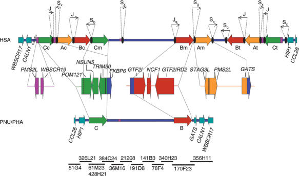
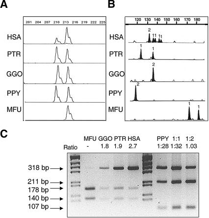
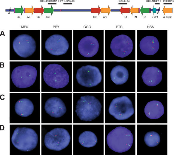
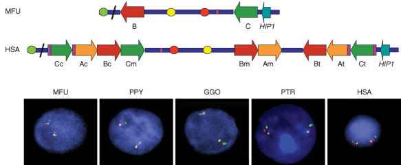
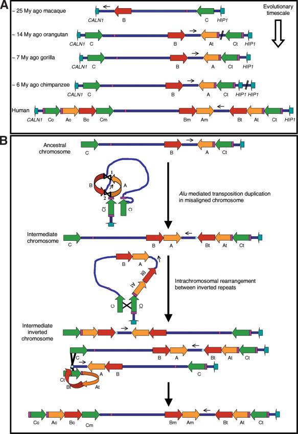
About 5% of the human genome consists of segmental duplications or low-copy repeats, which are large, highly homologous (>95%) fragments of sequence. It has been estimated that these segmental duplications emerged during the past approximately 35 million years (Myr) of human evolution and that they correlate with chromosomal rearrangements. Williams-Beuren syndrome (WBS) is a segmental aneusomy syndrome that is the result of a frequent de novo deletion at 7q11.23, mediated by large (approximately 400-kb) region-specific complex segmental duplications composed of different blocks. We have precisely defined the structure of the segmental duplications on human 7q11.23 and characterized the copy number and structure of the orthologous regions in other primates (macaque, orangutan, gorilla, and chimpanzee). Our data indicate a recent origin and rapid evolution of the 7q11.23 segmental duplications, starting before the diversification of hominoids (approximately 12-16 million years ago [Mya]), with species-specific duplications and intrachromosomal rearrangements that lead to significant differences among those genomes. Alu sequences are located at most edges of the large hominoid-specific segmental duplications, suggesting that they might have facilitated evolutionary rearrangements. We propose a mechanistic model based on Alu-mediated duplicated transposition along with nonallelic homologous recombination for the generation and local expansion of the segmental duplications. The extraordinary rate of evolutionary turnover of this region, rich in segmental duplications, results in important genomic variation among hominoid species, which could be of functional relevance and predispose to disease.


Figures

Figure 1.
Figure 1.
Schematic representation of the genomic structure of the WBS deletion region with the flanking segmental duplications in humans (HSA), and the homologous region in baboon (PNU/PHA). The large blocks of segmental duplications in the human map (A in yellow, B in red, C in green) are represented by thick arrows to indicate their relative orientation with respect to each other. They are exclusively present in the human map, whereas the baboon's genome contains the ancestral loci as single-copy and no large segmental duplication. The blue line represents the single-copy region, and the genes located immediately outside the region in both directions are represented as light blue arrows indicating the transcriptional direction. Some of the multiple-copy modules present in other chromosome 7 locations are shown in purple. The composition of each duplicated block with the corresponding transcriptional units is shown below the human map. Black ovals represent the Alu repeats located at the edges of the segmental duplications in the human map, with arrows indicating their orientation and approximate size (either partialor full Alu elements) shown on top. Note that the entire region including the ancestral loci of the segmental duplications is inverted in baboon with respect to the flanking genes. To define the baboon genomic structure, a clone contig with sequenced BACs from the RP41 library available in public databases has been assembled (NISC Comparative Sequencing Initiative), shown at the bottom.
Figure 2.
Figure 2.
Representative assays of copy number quantification in human (HSA), chimpanzee (PTR), gorilla (GGO), orangutan (PPY), and macaque (MFU) DNA, by comparison of paralogous sequence variants (PSVs) and microsatellites located in the segmental duplications. (A) A deletion/insertion PSV in block A distinguishes the ancestral STAG3 gene copy with respect to the pseudogene copies L1, L2, and L3. The STAG3/STAG3L copy ratio calculated for each species was: 0.47 ± 0.1 in HSA, 1.09 ± 0.1 in PTR, 1.04 ± 0.06 in GGO, and 0.95 ± 0.1 in PPY. Numbers on top show the amplimer size (in bp). (B) A microsatellite located between NCF1 and GTF2I in block B (BBSTR1, Bayés et al. 2003). All nonhuman primates displayed one or two alleles indicative of a single locus, whereas humans revealed six alleles corresponding to three different loci. The number of inferred alleles is indicated over each peak. (C)A restriction assay for a PSV of the TRIM50 gene in block C. In all primates but orangutan, there was a differential restriction site for NgoMIV. In orangutan, another assay with MluNI was performed and compared with artificial situations displaying 1:1, 2:1 ratios. Ratios between restriction products are shown at the top of each sample.
Figure 3.
Figure 3.
Number of blocks of segmental duplications detected by interphase FISH in the different primates with selected human BAC clones as probes. The location of all probes with respect to a representation of the human map is shown on top. (A) One single signal per chromosome in MFU, two in PPY, GGO, and PTR and four in HSA are detected with CITBI-E1-2601G15 (block A, 7q22, green), whereas CTB-139P11 (HIP1 locus, red) is single-copy in all species except chimpanzee, where it is duplicated. (B) In each species BAC RP11-204E14 (block B, green) displays one signal per chromosome except for humans, where three signals are found. (C) A single signal per chromosome is found in all the nuclei with RP11-622P13 (STX1A locus, red), whereas CTD-2528D12 (block C, green) displays one signal per chromosome in MFU, but two in PPY, GGO, and PTR and three in HSA. The single-copy STX1A locus is located in between the two (hominoids) or three (humans) signals of block C sequences. (D) CTB-139P11 (HIP1 locus, green) shows one signal per chromosome in all interphase nuclei except for PTR, which shows two signals indicating a duplication. Both copies of the HIP1 locus are located telomeric to the STX1A locus (RP11-622P13, red).
Figure 4.
Figure 4.
Genomic organization and gene order in the different species by multicolor FISH. Interphase FISH with three probes: BAC RP11-421B22 (CALN1 locus, green), BAC RP11-622P13 (STX1A locus, red), and PAC RP4-665P05 (GTF2IRD1 locus, yellow) showed the relative organization of the two loci within the WBS critical region with respect to an outside gene. A regional inversion is found in all primates but macaque, whose genomic structure is therefore identical to that of baboon and mouse. The location of each probe with respect to the human and predicted macaque maps is shown at the top.
Figure 5.
Figure 5.
Phylogenetic trees based on the neighbor-joining method (nucleotide gamma: Tamura Nei) or Bayesian method. Subindexes c, m, t, and 7q22 refer to centromeric, medial, telomeric, and 7q22 chromosomal localization of the human PSVs, respectively. Number 1 refers to the copy more similar to the ancestral human gene, and number 2 to the other copy. (A) Neighbor-joining tree obtained for the STAG3 gene PCR product (BA/STAG3) including HSA, PTR, GGO, PPY, MFU, and PNU. Branch numbers refer to the neighbor-joining bootstrap values/clade credibility values for the Bayesian tree, which was of identical conformation. (B) Bayesian tree obtained for the Block C Large fragment, including HSA, PTR, and PNU. Branch values refer to the clade credibility values.
Figure 6.
Figure 6.
Genomic structure of the orthologous region to human 7q11.23 in the different primates, and hypothetical model for the sequential evolution of the region. (A) Schematic representation of the chromosome region in each primate species. A first inversion of the WBS region must have occurred in an ancestral chromosome to all hominoids. The orangutan and gorilla chromosomes appear identical except for the absence of the block C-block A junction in orangutan, whereas gorilla and chimpanzee chromosomes are identical except for the segmental duplication containing the HIP1 gene in chimpanzee. (B) Predicted human lineage-specific rearrangements from a hypothetical ancestral chromosome identical to that of gorilla. A unique complex intrachromosomal rearrangement from the ancestral chromosome created an intermediate chromosomal structure by two shuffling events between Alu elements, represented as 1 and 2 indicating the order of occurrence. By a similar mechanism of Alu-mediated duplicative transposition, the chimpanzee chromosome could have been generated (data not shown), with a duplication of the HIP1 containing block instead. A putative intrachromosomal paracentric inversion in the intermediate chromosome could have been mediated by the blocks C, which are flanking the region in inverted orientation. Interchromosomal NAHR in an inversion carrier of this intermediate chromosome could have led to duplication of the entire segmental duplication-containing blocks C, A, and B onto the centromeric position. The presence of Alu elements located at the edges of the blocks suggests Alu-mediated genome shuffling in these final steps of the generation of large segmental duplications.

Similar articles

Cited by

References

    1. Altschul, S.F., Gish, W., Miller, W., Myers, E.W., and Lipman, D.J. 1990. Basic local alignment search tool. J. Mol. Biol. 215: 403-410. - PubMed
    1. Armengol, L., Pujana, M.A., Cheung, J., Scherer, S.W., and Estivill, X. 2003. Enrichment of segmental duplications in regions of breaks of synteny between the human and mouse genomes suggest their involvement in evolutionary rearrangements. Hum. Mol. Genet. 12: 2201-2208. - PubMed
    1. Babcock, M., Pavlicek, A., Spiteri, E., Kashork, C.D., Ioshikhes, I., Shaffer, L.G., Jurka, J., and Morrow, B.E. 2003. Shuffling of genes within low-copy repeats on 22q11 (LCR22) by Alu-mediated recombination events during evolution. Genome Res. 13: 2519-2532. - PMC - PubMed
    1. Bailey, J.A., Liu, G., and Eichler, E.E. 2003. An Alu transposition model for the origin and expansion of human segmental duplications. Am. J. Hum. Genet. 73: 823-834. - PMC - PubMed
    1. Batzer, M.A. and Deininger, P.L. 2002. Alu repeats and human genomic diversity. Nat. Rev. Genet. 3: 370-379. - PubMed

WEB SITE REFERENCES

    1. http://www.ncbi.nlm.nih.gov/; NCBI home page.
    1. http://www.nisc.nih.gov/; The NIH Intramural Sequencing Center (NISC). - PubMed
    1. http://www.ensembl.org/; Ensembl Genome Browser.
    1. http://www.genome.ucsc.edu/; UCSC Genome Browser Home.
    1. http://frodo.wi.mit.edu/cgi-bin/primer3/primer3_www.cgi; Primer3 Input.

Publication types

Associated data

LinkOut - more resources