. 2005 Sep 13;102(37):13212-7.
doi: 10.1073/pnas.0506306102. Epub 2005 Sep 1.

A lentiviral microRNA-based system for single-copy polymerase II-regulated RNA interference in mammalian cells

Affiliations

A lentiviral microRNA-based system for single-copy polymerase II-regulated RNA interference in mammalian cells

Frank Stegmeier et al. Proc Natl Acad Sci U S A. .

Abstract

The advent of RNA interference has led to the ability to interfere with gene expression and greatly expanded our ability to perform genetic screens in mammalian cells. The expression of short hairpin RNA (shRNA) from polymerase III promoters can be encoded in transgenes and used to produce small interfering RNAs that down-regulate specific genes. In this study, we show that polymerase II-transcribed shRNAs display very efficient knockdown of gene expression when the shRNA is embedded in a microRNA context. Importantly, our shRNA expression system [called PRIME (potent RNA interference using microRNA expression) vectors] allows for the multicistronic cotranscription of a reporter gene, thereby facilitating the tracking of shRNA production in individual cells. Based on this system, we developed a series of lentiviral vectors that display tetracycline-responsive knockdown of gene expression at single copy. The high penetrance of these vectors will facilitate genomewide loss-of-function screens and is an important step toward using bar-coding strategies to follow loss of specific sequences in complex populations.


Figures
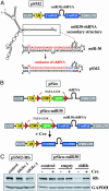
Fig. 1.
Fig. 1.
Pol II-driven miR30-based shRNAs function efficiently at single copy. (A) Schematic representation of the pSM2 vector structure (see Fig. 6 for additional information), showing the secondary structure fold of the miR30-based shRNAs as predicted by rnafold (http://rna.tbi.univie.ac.at/cgi-bin/RNAfold.cgi). In pSM2, the region coding for the mature miR30 miRNA (upper hairpin) is replaced with sequences that encode shRNAs targeting any gene of choice (shRNA, lower hairpin). The antisense strand, which encodes the targeting siRNA is shown in red. (B) Schematic representation of pSico and pSico–miR30 before and after Cre-mediated recombination. (C) U2OS cells were transduced at the indicated MOIs with pSM2 containing an shRNA that targets the Rb transcript (pSM2–Rb) and cultured for 1 week in puromycin (1 μg/ml)-containing medium. For pSico–miR30, U2OS cells were infected with the indicated lentivirus (MOI <0.3) and sorted for GFP-positive cells. These cells were infected with Ad–Cre and sorted for GFP-negative cells 4 days postinfection. Cell extracts were immunoblotted for Rb and GAPDH (loading control) protein levels. Uninfected cells serve as control.
Fig. 2.
Fig. 2.
Tet-regulatable pPRIME vectors generate highly penetrant knockdown at single copy. (A) Schematic representation of the TREX system. In the absence of DOX, the Tet repressor (TetR) binds to the Tet-operator binding sites located downstream of CMV's TATA box, thereby preventing transcription. Binding of DOX to TetR causes its displacement from the CMV promoter, thus allowing for transcription of the GFP–miR30–shRNA transcript. (B) 293 TREX cells were transduced with the indicated lentiviruses (MOI <0.4), grown in DOX for 3–4 days, and sorted for GFP-positive cells. Cells were split and grown either in the presence (+DOX) or absence (-DOX) of DOX for 1 week. Whole-cell extracts were immunoblotted for Rb, GFP, and GAPDH (loading control). (C) 293 TREX cells were transduced with the indicated lentiviruses. Cells infected at low MOI (<0.4) and high MOI (MOI = 5) were grown in DOX for 3–4 days and sorted for GFP-positive cells. Cells infected at the high MOI were also sorted for the highest 10% of GFP expression levels. Whole-cell extracts were immunoblotted against Rb, GFP, and GAPDH (loading control). (D) Schematic representation of the Tet-ON system. In the absence of DOX, the reverse tTA (rtTA) does not bind to the TET promoter. Binding of DOX to rtTA promotes its binding to the TET promoter, thereby initiating the transcription of the GFP–miR30–shRNA transcript. (E) U2OS Tet-ON cells were transduced with the indicated lentiviruses (MOI <0.4), grown in DOX for 3–4 days, and sorted for GFP-positive cells. Cells were treated and analyzed as described in B.(F) Images of 293 TREX transduced with pPRIME–TREX–GFP–Rb grown either in the absence (Lower) of DOX or the presence of DOX (induced for 16 h, Upper). (G) Schematic representation of the Tet-OFF system. In the absence of DOX, the tTA is bound to the TET promoter and drives transcription of the GFP–miR30–shRNA transcript. Binding of DOX to tTA leads to its displacement from the promoter and shutdown of transcription. (H and I) HeLa Tet-OFF and U2OS Tet-OFF cells were transduced with the indicated lentiviruses (MOI <0.4), treated, and analyzed as described in B.
Fig. 3.
Fig. 3.
Kinetics and dose–responsiveness of Rb regulation by pPRIME–TET–GFP–Rb. (A) Clonal isolates of the HeLa Tet-OFF cells expressing pPRIME–Tet–GFP–Rb (see Fig. 2H) were grown under inducing conditions (-DOX) and immunoblotted for Rb, GAPDH, and GFP protein levels. (B) Tet-OFF clone 1 (see A) expressing GFP–miR30–Rb was cultured for 1 week in medium containing the indicated concentration of DOX, and cell extracts were blotted for the indicated proteins. (C) Tet-OFF clone 1 was cultured for 1 week in the presence of 1 μg/ml DOX (repressing condition). At day 0, the drug was withdrawn from the culture medium, and cells were harvested at the indicated time points. Whole-cell extracts were analyzed by Western blotting for the indicated proteins. (D) Tet-OFF clone 1 was cultured for 1 week under inducing conditions (-DOX), followed by addition of DOX at a concentration of 1 μg/ml. Cells were harvested just before DOX treatment (day 0) and at the indicated time points. Whole-cell extracts were analyzed by Western blotting for the indicated proteins.
Fig. 4.
Fig. 4.
pPRIME vectors can accommodate a variety of reporter genes. (A) Schematic representation of pPRIME–CMV derivatives. For more detailed vector information see Fig. 6. PGK, phosphoglycerate kinase promoter; IRES, internal ribosome entry site. (B) U2OS cells were transduced with the indicated lentiviruses (MOI <0.4, uninfected cells served as control). GFP-, dsRed-, and LNGFR-expressing cells were FACS-sorted 4 days after infection, and Neo-expressing cells (CMV, CMV-NEO, and CMV-GIN) were selected with 500 μg/ml G418 for 1 week. Drug selection was withdrawn 1.5 days before harvesting the cells. Whole-cell extracts were analyzed by immunoblotting for the indicated proteins.

References

    1. Hannon, G. J. & Rossi, J. J. (2004) Nature 431, 371-378. - PubMed
    1. Silva, J., Chang, K., Hannon, G. J. & Rivas, F. V. (2004) Oncogene 23, 8401-8409. - PubMed
    1. Dykxhoorn, D. M., Novina, C. D. & Sharp, P. A. (2003) Nat. Rev. Mol. Cell Biol. 4, 457-467. - PubMed
    1. Elbashir, S. M., Harborth, J., Lendeckel, W., Yalcin, A., Weber, K. & Tuschl, T. (2001) Nature 411, 494-498. - PubMed
    1. Mittal, V. (2004) Nat. Rev. Genet. 5, 355-365. - PubMed

Publication types