. 2005 Sep 13;102(37):13236-41.
doi: 10.1073/pnas.0506218102. Epub 2005 Sep 1.

Knockdown of Pu.1 by small interfering RNA in CD34+ embryoid body cells derived from mouse ES cells turns cell fate determination to pro-B cells

Affiliations

Knockdown of Pu.1 by small interfering RNA in CD34+ embryoid body cells derived from mouse ES cells turns cell fate determination to pro-B cells

Gang-Ming Zou et al. Proc Natl Acad Sci U S A. .

Abstract

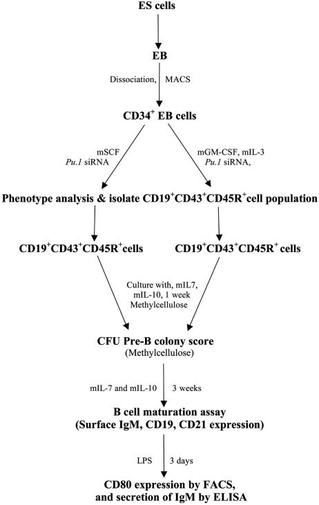
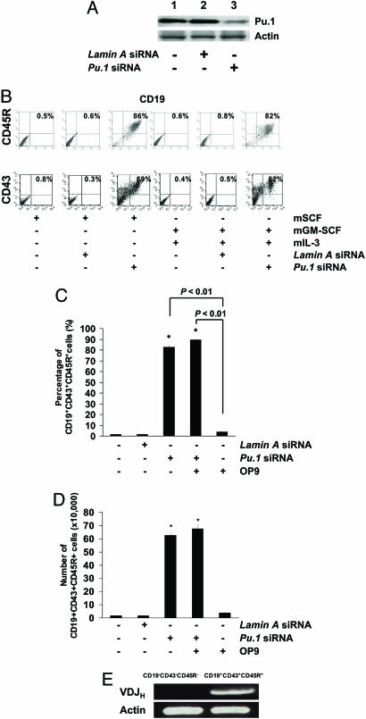
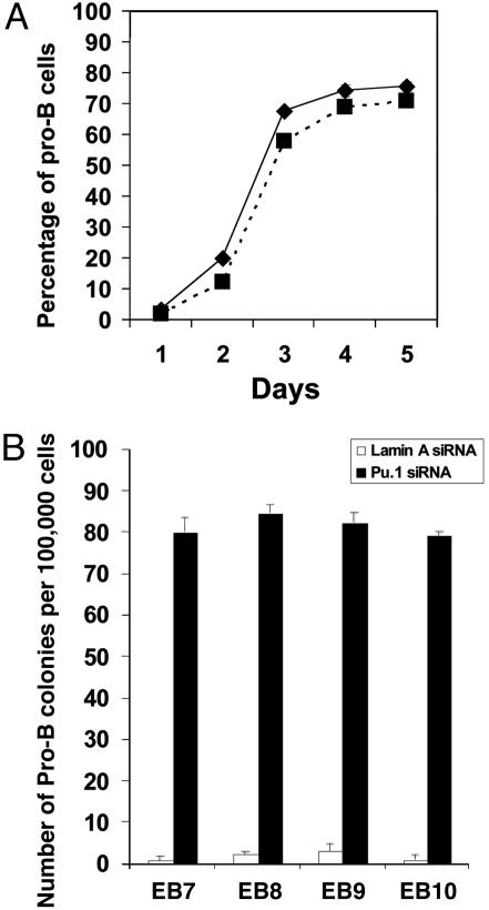
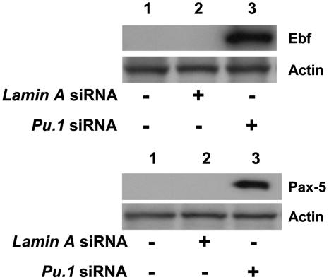
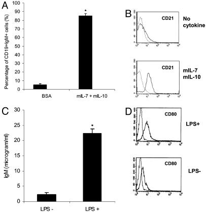
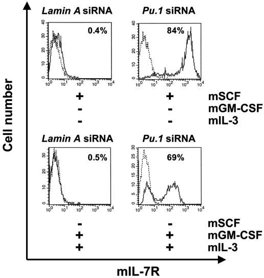
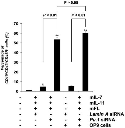
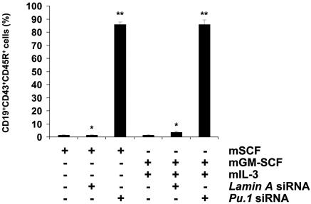
The factors that regulate murine ES cell-derived hematopoietic progenitor cell (HPC) commitment to the B lymphocyte lineage remain unclear. Pu.1 plays an essential role in the development of all lymphoid lineages; however, it also regulates commitment to other blood cell lineages. In this study, we found evidence for early B cell lineage commitment as determined by coexpression of CD19 and CD45R (B220) when Pu.1 expression was knocked down in HPC by specific small interfering RNA (siRNA); moreover, the expression of early B cell factor (Ebf) and paired box protein 5 (Pax-5) transcription factors was induced when cells were treated by Pu.1 siRNA, but not by control siRNA. We also found that siRNA-mediated knockdown of Pu.1 expression was more efficient in generating progenitor B cells (pro-B cells) compared with the more common in vitro method of B lymphoid development by means of coculture of CD34+ embryoid body (EB) cells with OP9 stromal cells. To investigate whether this phenomenon also exists in HPC from other sources, we then knocked down Pu.1 gene expression in CD34+ murine bone marrow cells and found a similar effect of increased production of CD19+CD43+CD45R+ progenitor B cells upon the siRNA-mediated decrease in Pu.1 expression. We conclude that, in early B cell development from ES cell-derived HPC, constitutive Pu.1 expression inhibits the earliest B cell development through repressing early B cell factor and paired box protein 5 expression, although lower levels of Pu.1 expression in HPC play a key role in promoting B cell fate determination.


Figures

Fig. 1.
Fig. 1.
General outline of this protocol. ES cells were differentiated into EBs in the absence of murine leukemia inhibitory factor. CD34+ cell populations were isolated from whole EB cell suspension by magnetic associated cell sorting magnetic bead cell separation. Cells were then transfected with Pu.1 siRNA and maintained with mSCF in culture, or secondary differentiation was initiated in myeloid differentiation conditions; i.e., mGM-CSF and mIL-3 were added in the medium. Pro-B cell formation was examined in the CD19+CD43+CD45R+ phenotype. To test whether the B progenitors were functional, cells were matured and examined for CD21 expression, and further examined for their LPS stimulation response, such as CD80 expression and IgM secretion.
Fig. 2.
Fig. 2.
Knockdown of Pu.1 by siRNA leads to CD19+CD43+CD45R+ pro-B cells. (A) Western blot analysis was performed to confirm the effect of Pu.1 siRNA on the target gene level of expression. As shown, Pu.1 expression was reduced when cells were treated by Pu.1 siRNA (lane 3) compared with siRNA untreated cells (lane 1) or control (Lamin A) siRNA-treated cells for 72 h. Data are representative of one of three individual experiments. (B) Pu.1 siRNA-transfected CD34+ cells maintained with SCF differentiated to CD19+CD43+CD45R+ pro-B cells. (C) Pu.1 siRNA-transfected CD34+ cells cultured with mGM-CSF and mIL-3 differentiated into CD19+CD43+CD45R+ pro-B cells. Data from one representative experiment from three individual experiments is shown. (D) Comparison of pro-B cells generated between siRNA-mediated Pu.1 knockdown of gene expression compared with the OP9 coculture system. Knockdown of Pu.1 expression induced over 80% of the CD34+ cells to become pro-B cells within 72 h. However, simple coculture of CD34+ EB cells with OP9 stromal cells induced only 5% of cells to become pro-B cells in the same time period. Combination of knockdown of Pu.1 expression by means of siRNA treatment with coculture with OP9 cells could not significantly increase the frequency of pro-B cells compared with the siRNA alone group. (E) Generated CD19+CD43+CD45R+ cells express VDJ gene rearrangement examined by RT-PCR.
Fig. 3.
Fig. 3.
Kinetics in pro-B cell production from Pu.1 siRNA-treated CD34+ EB cells, and a comparison of the potential for pro-B cell development of these cells from different EB ages. (A) After Pu.1 siRNA treatment, in day 1, <5% of cells are pro-B cells; in day 2, it increased to ≈20%; on day 3, the percentage of pro-B cells has increased to 60–70%; then the percentage of pro-B cells gradually increased until day 5 (solid line, cells cultured with mSCF; dotted line, cells cultured with mGM-CSF and mIL-3). (B) CD34+ EB cells from different age EBs have similar potential in pro-B cell development under the Pu.1 siRNA treatment.
Fig. 4.
Fig. 4.
CFU-pre-B colony development from a CD19+CD43+CD45R+ cell populations (open bars, CD19-CD43-CD45R- cells; filled bars, CD19+CD43+CD45R+ cells). In the control group, without the stimulation by cytokines mIL-7 and mIL-10, there was no pre-B colony formation. When CD19+CD43+CD45R+ cells were treated with mIL-7, some pre-B colonies formed; however, when mIL-7 and mIL-10 were combined, the frequency of CFU-pre-B cells increased dramatically (*, P < 0.05 for CD19+CD43+CD45R+ cells versus CD19-CD43-CD45R- cells in mIL-7 culture; **, P < 0.01 for CD19+CD43+CD45R+ cells versus CD19-CD43-CD45R- cells in mIL-7 plus mIL-10 culture).
Fig. 5.
Fig. 5.
Knockdown of Pu.1 expression in CD34+ cells induces both Ebf and Pax-5 expression. (Upper) Ebf expression appears in Pu.1 siRNA-treated CD34+ EB cells primed by mGM-CSF and mIL-3 treatment for 72 h. Lane 1, untreated cells; lane 2, cells treated with control Lamin A siRNA; lane 3, cells treated with Pu.1 siRNA. Representative data from one of three individual experiments are shown. (Lower) Pax-5 expression is increased in Pu.1 siRNA-treated CD34+ EB cells. Lane 1, untreated cells; lane 2, cells treated with control Lamin A siRNA; lane 3, cells treated with Pu.1 siRNA. Representative data from one of three individual experiments are shown.
Fig. 6.
Fig. 6.
Pro-B cell can be further differentiated to mature B cells. (A) Frequency of functional B cell (CD19+IgM+) formation from CD19+CD43+CD45R+ pro-B cells. (B) CD21 expression analysis by flow cytometry. Cytokine stimulation (mIL-7 and mIL-10) is able to up-regulate CD21 expression in these B cells. Representative data from one of three individual experiments are shown. (C) sIgM assay by ELISA on CD19+CD43+CD45R+ cells stimulated by LPS. Data are the mean ± SD of three independent experiments. Lane 1, untreated cells; lane 2, cells treated with LPS. (D) LPS up-regulates CD80 expression in the CD19+CD43+CD45R+ cell population (*, P < 0.01).
Fig. 7.
Fig. 7.
Knockdown of Pu.1 expression results in mIL-7R expression on CD34+ EB cells cultured either in maintenance conditions (mSCF alone) or myeloid differentiation conditions (mGM-CSF and mIL-3). (Upper) CD34+ EB cells cultured with mSCF did not express mIL-7R when treated with control siRNA; however, upon treatment with Pu.1 siRNA, mIL-7R expression was induced and detected by flow cytometry. Representative data of one of three individual experiments are shown. (Lower) In CD34+ EB cells cultured with mGM-CSF and mIL-3, mIL-7R expression was also induced when cells were treated by Pu.1 siRNA, but not control siRNA. Representative data from one of three individual experiments are shown.
Fig. 8.
Fig. 8.
Knockdown of Pu.1 expression on CD34+ EB cells enhances the frequency of CD19+CD43+CD45R+ cell production mediated by mIL-7, mIL-11, and mFL in vitro. CD34+ EB cells were transfected with either Lamin A siRNA (control) or Pu.1 siRNA (50 nM), and cultured with or without OP9 cells and with or without cytokines mIL-7, mIL-11, and mFL for 72 h. Cells were then collected and analyzed by flow cytometry for cell surface marker expression. Data are the mean ± SD of three independent experiments. Pu.1 siRNA-treated CD34+ cells showed a 10-fold increase in the production of CD19+CD43+CD45R+ cells compared with the cells treated with control siRNA (*, P < 0.01).
Fig. 9.
Fig. 9.
Knockdown of Pu.1 expression in CD34+ murine bone marrow cells enhances cell fate determination to pro-B cells. CD34+ marrow cells were transfected with control siRNA or Pu.1 siRNA and then cultured with mSCF or mGM-CSF plus mIL-3 for 72 h, before harvesting for FACS analysis. CD19, CD43, and CD45R expression was significantly induced in these bone marrow hematopoietic cells when they were treated by Pu.1 siRNA for 72 h. Data are the mean ± SD of three independent experiments.

References

    1. Keller, G., Lacaud, G. & Robertson, S. (1999) Exp. Hematol. 27, 777-787. - PubMed
    1. Daley, G. Q. (2003) Ann. N.Y. Acad. Sci. 996, 122-133. - PubMed
    1. Hamaguchi-Tsuru, E., Nobumoto, A., Hirose, N., Kataoka, S., Fujikawa-Adachi, K., Furuya, M. & Tominaga, A. (2004) Br. J. Haematol. 124, 819-827. - PubMed
    1. Lieber, J. G., Webb, S., Suratt, B. T., Young, S. K., Johnson, G. L., Keller, G. M. & Worthen, G. S. (2004) Blood 103, 852-859. - PubMed
    1. Fujimoto, T. T., Kohata, S., Suzuki, H., Miyazaki, H. & Fujimura, K. (2003) Blood 102, 4044-4051. - PubMed

Publication types