. 2005 Oct;37(10):1047-54.
doi: 10.1038/ng1634. Epub 2005 Sep 4.

The melanocyte differentiation program predisposes to metastasis after neoplastic transformation

Affiliations

The melanocyte differentiation program predisposes to metastasis after neoplastic transformation

Piyush B Gupta et al. Nat Genet. 2005 Oct.

Abstract

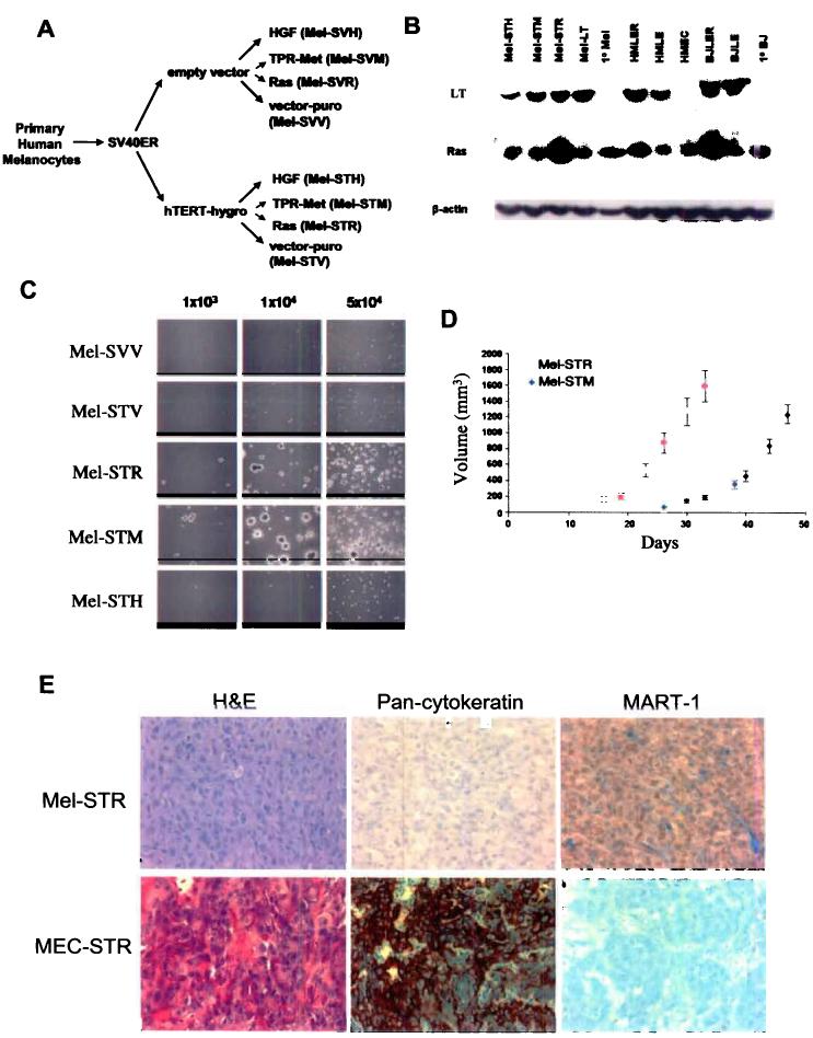
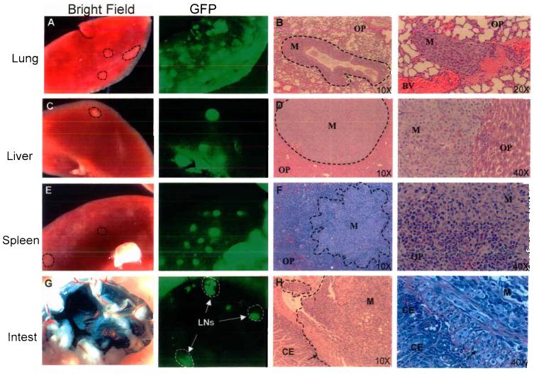
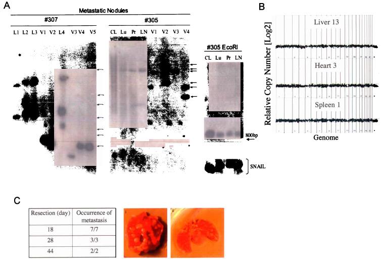
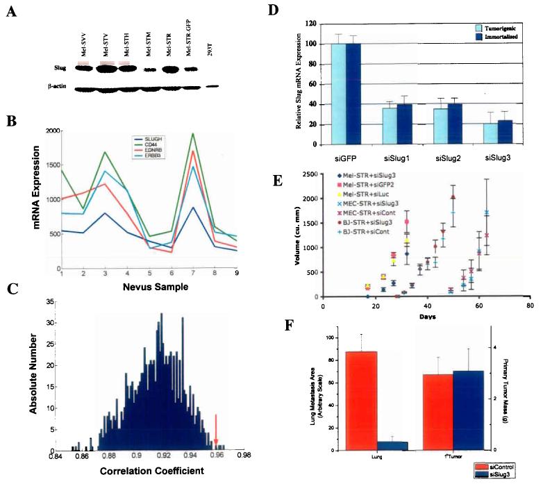
The aggressive clinical behavior of melanoma suggests that the developmental origins of melanocytes in the neural crest might be relevant to their metastatic propensity. Here we show that primary human melanocytes, transformed using a specific set of introduced genes, form melanomas that frequently metastasize to multiple secondary sites, whereas human fibroblasts and epithelial cells transformed using an identical set of genes generate primary tumors that rarely do so. Notably, these melanomas have a metastasis spectrum similar to that observed in humans with melanoma. These observations indicate that part of the metastatic proclivity of melanoma is attributable to lineage-specific factors expressed in melanocytes and not in other cell types analyzed. Analysis of microarray data from human nevi shows that the expression pattern of Slug, a master regulator of neural crest cell specification and migration, correlates with those of other genes that are important for neural crest cell migrations during development. Moreover, Slug is required for the metastasis of the transformed melanoma cells. These findings indicate that melanocyte-specific factors present before neoplastic transformation can have a pivotal role in governing melanoma progression.


Figures

Figure 1
Figure 1
Characterization of retrovirus-transduced primary human melanocytes. a, Schematic of the transformation protocol. b, Expression of the LT, Ras, and TPR-met proteins in primary and engineered cell lines. c, Anchorage-independent colony formation of engineered melanocyte (Mel) cell lines requires expression of the SV40ER and hTERT, together with either RasV12 or TPR-met. Assays were performed in triplicate for each cell line shown and a representative field photographed. d, Subcutaneous tumor growth of engineered melanoma cell lines in athymic nude mice in vivo (n>=12). Error bars reflect standard errors. e, Hematoxylin & eosin staining, and MART-1 and pan-cytokeratin immunostainings of histological sections prepared from engineered subcutaneous mammary epithelial cell (MEC-STR) and melanoma (Mel-STR) tumors.
Figure 2
Figure 2
Primary Mel-STR melanomas give rise to widespread metastases in vivo. Immunocompromised NOD/SCID mice were injected subcutaneously with 5×10⁵ GFP-labeled Mel-STR cells. Organs were harvested at necropsy and were immediately visualized for GFP epifluorescence, or fixed for subsequent paraffin embedding. Images of whole-organs (a,c,e,g) and H&E-stained sections (b,d,f,h) were captured of lungs, livers, spleens, and small intestines taken from representative metastasis-bearing mice. Dashed lines demarcate tumor cell regions. M, metastasis; OP, organ parenchyma; CE, colonic epithelium; BV, blood vessel; LN, lymph node; Intest, small bowel. Arrows in h indicate a front of melanoma cells invading from the outside surface of the small bowel, through the muscularis propria, into the colonic epithelium.
Figure 3
Figure 3
Primary Mel-STR melanomas rapidly seed distinct metastatic clones to secondary organs. a, Southern blot analysis of the metastases arising in single Mel-STR-injected mice. Lanes were loaded with BamHI-cleaved genomic DNA extracted either from organs (#305Lu, Pr, LN), or from the parental Mel-STR cell line (Parental, #305CL) immediately prior to injection, or from Mel-STR cells that were isolated from individual metastatic nodules in vivo and expanded in culture (L1-4, V1-5). Arrows indicate independent insertion sites. BamHI cleaves once within the vector provirus, upstream of the probed retroviral GFP sequence. GFP probe specificity in lanes exhibiting smears was confirmed by digesting the extracted DNAs with EcoRI, which cleaves twice within the vector provirus and collapses the smears into a single internal 880bp fragment independent of provirus integration site. Genomic integrity in samples exhibiting smears was also confirmed by probing EcoRI-digested DNA for an endogenous 1 kb region upstream of the SNAIL gene, revealing a single intact 10.7 kb band without detectable degradation. HindIII digestion of the extracted DNAs recapitulated the results observed with BamHI cleavage (data not shown). L, lung; V, liver; LN, lymph node; CL, parental Mel-STR cell line; Pr, primary tumor lysate; Lu, whole-lung lysate. b, Whole genome array CGH performed on DNA isolated from metastatic nodules isolated from the liver, spleen, and heart of a single metastasis-laden animal. No significant genome copy number alterations are observed in any of the metastatic nodules relative to the parental primary tumor obtained from the same animal. Data are displayed as log2(CY3/CY5 intensity ratios) from pter to qter with chromosome 1 on the left and chromosomes 22 and X on the right. c, Mice were injected subcutaneously with Mel-STR cells and the primary melanomas resected at the indicated times. All mice were sacrificed at 48 days post-injection. The fraction of mice with visible lung metastases upon necropsy is indicated. (i, ii) are representative melanoma-burdened lungs from mice whose primary melanomas were excised at day 18 or day 28 after injection, respectively. Arrows indicate individual metastatic nodules.
Figure 4
Figure 4
Suppression of Slug expression inhibits melanoma metastasis in vivo. a, Western blot analysis of Slug protein expression. Slug protein levels were examined in melanocyte cell lines engineered with LT (SVV), LT, hTERT & HGF (STH), LT, hTERT & TPR-Met (STM), LT, hTERT & RasV12 (STR), or LT, hTERT, RasV12 & GFP (STR-GFP), and in transformed kidney cells (293T). b, mRNA expression levels as determined by microarray analysis. Transcript levels of SLUGH, CD44, EDNRB and ERBB3 were correlated in 9 human nevus samples. c, Reference statistics for CD44 (35). 1000 correlation coefficients were obtained by randomly permuting the SLUGH expression profile template. Shown is a histogram plot of correlations corresponding to the rank of CD44 in the SLUGH neighborhood list. The correlation coefficient of CD44 with the non-permuted SLUGH template is indicated by a red arrow (0.958). There are only 4 permuted correlations larger than this value, resulting in strong statistical significance (p=0.004). d, Quantitative RT-PCR performed on immortalized and transformed melanocyte populations stably expressing either siSlug (siSlug1, siSlug2, siSlug3) or control (siGFP2) siRNAs. Data were normalized to GAPDH expression and plotted as a percentage relative to Slug transcript levels in siGFP2-expressing cells. e, In vivo tumor growth curves of Mel-STR, MEC-STR, and BJ-STR cells stably expressing either siSlug3 or control (siGFP2, siLuc) siRNA contructs. f, Quantification of lung metastasis burden in siSlug3- or control siRNA (siGFP2, siLuc)- Mel-STR melanoma lines. Random lung sections were stained with a human-specific vimentin antibody and the area of brown color corresponding to stained Mel-STR cells was quantified using NIH Image software (35).

References

    1. Lens MB, Dawes M. Global perspectives of contemporary epidemiological trends of cutaneous malignant melanoma. Br J Dermatol. 2004;150:179–85. - PubMed
    1. Beddingfield FC., 3rd The melanoma epidemic: res ipsa loquitur. Oncologist. 2003;8:459–65. - PubMed
    1. Bartkova J, et al. The p16-cyclin D/Cdk4-pRb pathway as a functional unit frequently altered in melanoma pathogenesis. Cancer Res. 1996;56:5475–83. - PubMed
    1. Omholt K, Platz A, Kanter L, Ringborg U, Hansson J. NRAS and BRAF mutations arise early during melanoma pathogenesis and are preserved throughout tumor progression. Clin Cancer Res. 2003;9:6483–8. - PubMed
    1. Chin L. The genetics of malignant melanoma: lessons from mouse and man. Nat Rev Cancer. 2003;3:559–70. - PubMed

Publication types

MeSH terms