. 2005 Sep 14;127(36):12647-56.
doi: 10.1021/ja053254z.

The octarepeat domain of the prion protein binds Cu(II) with three distinct coordination modes at pH 7.4

Affiliations

The octarepeat domain of the prion protein binds Cu(II) with three distinct coordination modes at pH 7.4

Madhuri Chattopadhyay et al. J Am Chem Soc. .

Abstract

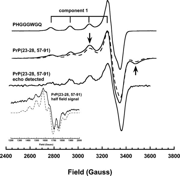
The prion protein (PrP) binds Cu2+ in its N-terminal octarepeat domain. This unusual domain is comprised of four or more tandem repeats of the fundamental sequence PHGGGWGQ. Previous work from our laboratories demonstrates that at full copper occupancy, each HGGGW segment binds a single Cu2+. However, several recent studies suggest that low copper occupancy favors different coordination modes, possibly involving imidazoles from histidines in adjacent octapeptide segments. This is investigated here using a combination of X-band EPR, S-band EPR, and ESEEM, along with a library of modified peptides designed to favor different coordination interactions. At pH 7.4, three distinct coordination modes are identified. Each mode is fully characterized to reveal a series of copper-dependent octarepeat domain structures. Multiple His coordination is clearly identified at low copper stoichiometry. In addition, EPR detected copper-copper interactions at full occupancy suggest that the octarepeat domain partially collapses, perhaps stabilizing this specific binding mode and facilitating cooperative copper uptake. This work provides the first complete characterization of all dominant copper coordination modes at pH 7.4.


Figures

Figure 1
Figure 1
X-band EPR spectra of PrP(23–28, 57–91) (200 μM) as a function of Cu2+ concentration, represented in equivalents. Three distinct species are observed, as can be clearly seen in the inset showing an expansion of the mI = −3/2 and mI = −½ hyperfine lines. The grids at the top identify the four hyperfine lines arising from coupling to the 63Cu (I = 3/2) nucleus for each spectral species. Spectra were obtained at approximately 77 K and νo = 9.44 GHz.
Figure 2
Figure 2
X-band EPR spectra showing the approximate equivalence of fully occupied PrP(23–28, 57–91) and PHGGGWGQ. The spectrum for PrP(23–28, 57–91) also reveals features associated with dipolar coupling (arrows). The echo detected spectrum of PrP(23–28, 57–91), obtained with a two pulse sequence (τ = 140 ns, νo = 9.63 GHz, T = 6 K), selects for slowly relaxing species and lacks the dipolar features, thus confirming a spectral superposition. The inset shows the half field signal (νo = 9.63 GHz) obtained from PrP(23–28, 57–91) with 2.0 equiv of Cu2+. The simulation (dashed line) for the g ≈ 2 spectrum was generated with a superposition of a mononuclear species (80%) and a coupled species (20%) using the parameters g = 2.227, g = 2.052, and A = 162 G and g = 2.176, g = 2.058, and A = 165 G, respectively, and a distance of 6.0 Å. The simulation of the half field signal (dashed line) used g = 2.174, g = 2.058, and A = 162 G and a distance of 4.9 Å. In addition, the g tensors for the Cu(II) ions were assumed to be collinear with an intercopper vector at a 10° ± 5° angle to the z direction.
Figure 3
Figure 3
X-band EPR spectra of PrP constructs containing N-methylated glycine (sarcosine, X) at specific positions to block component 1 binding. Methylation of the first or second glycine results in pure component 2 binding.
Figure 4
Figure 4
Three-pulse ESEEM spectra of constructs that exhibit component 2 binding showing single imidazole coordination. Spectra were obtained at 4.2 K from the g region of the spectrum with τ = 150 ns.
Figure 5
Figure 5
S-band EPR of HGXGW that favors component 2 binding. (A) Shows the full scan, (B) expansion of the mI = −½ line, and (C) the mI = −½ line from a sample containing 30% 17OH2. The five line multiplet at mI = −½ is consistent with coordination of two nitrogens. Insensitivity to 15N placement at the first and third glycines shows that these residues do not coordinate. Broadening by 17OH2 suggests that water contributes to the coordination sphere. Spectra (3.5 GHz) were acquired in D2O solution at 133 K.
Figure 6
Figure 6
Models of the three equatorial coordination modes. For component 3, X may represent either a fourth imidazole or a water molecule.
Figure 7
Figure 7
Three-pulse ESEEM comparing PrP(23–28, 57–91) with 4.0 and 1.0 equiv of Cu2+. At 4.0 equiv, component 1 dominates; the grid at the top shows the previously determined assignment. At 1.0 equiv, which favors component 3, the enhanced ΔmI = 2 feature at 4.1 MHz is consistent with multiple His coordination. The inset shows the S-band mI = −½ line obtained with 1.0 equiv. The seven- or nine-line pattern suggests 3N1O or 4N coordination by equivalent nitrogens.
Figure 8
Figure 8
Models of PrPC containing Cu2+ in the three coordination modes. The figure at upper left shows PrP(60–231) bound to a single Cu2+ with component 3 coordination. To the upper right, the copper binding octarepeat domain is expanded. In this mode, an additional His imidazole may also participate in copper binding. For components 1 and 2, only the octarepeat domains are shown.

Similar articles

Cited by

References

    1. Prusiner SB. Proc. Natl. Acad. Sci. U.S.A. 1998;95:13363–13383. - PMC - PubMed
    1. Prusiner SB. Science. 1997;278:245–251. - PubMed
    1. Legname G, Baskakov IV, Nguyen HO, Riesner D, Cohen FE, DeArmond SJ, Prusiner SB. Science. 2004;305:673–6. - PubMed
    1. Herms J, Tings T, Gall S, Madlung A, Giese A, Siebert H, Schürmann P, Windl O, Brose N, Kretzschmar H. J. Neurosci. 1999;19:8866–8875. - PMC - PubMed
    1. Vassallo N, Herms J. J. Neurochem. 2003;86:538–44. - PubMed

Publication types

MeSH terms