. 2005 Sep;43(9):4426-33.
doi: 10.1128/JCM.43.9.4426-4433.2005.

Detection of antibodies against adenovirus protein IX, fiber, and hexon in human sera by immunoblot assay

Affiliations

Detection of antibodies against adenovirus protein IX, fiber, and hexon in human sera by immunoblot assay

Ulrike Bauer et al. J Clin Microbiol. 2005 Sep.

Abstract

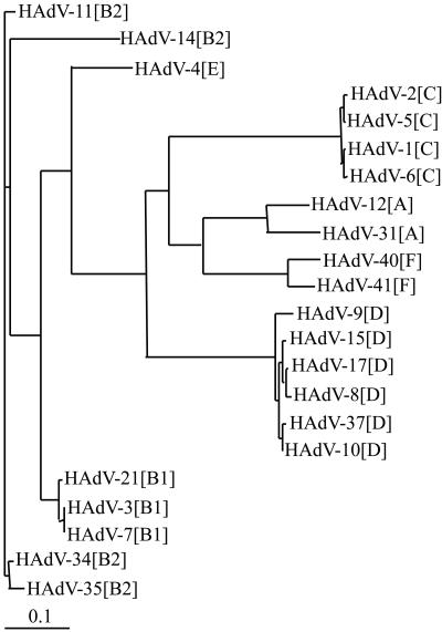
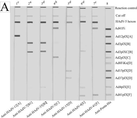
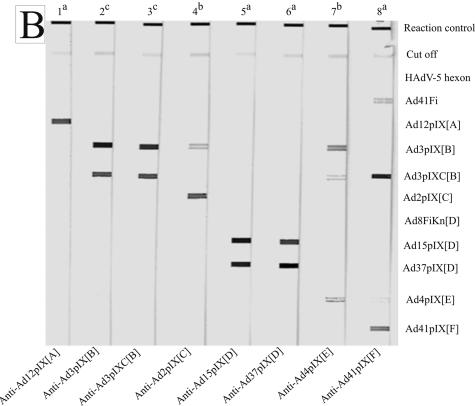
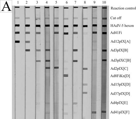
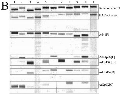
The 51 serotypes of human adenoviruses (HAdVs) of the genus Mastadenovirus are classified into the six species HAdV-A to HAdV-F. For the detection of genus- and species-specific antibodies in human sera an immunoblot assay was developed. The recombinant long fiber of HAdV-41[F] (Ad41Fi) and the native hexon of HAdV-5[C] were used as genus-specific antigens. The recombinant capsid protein IX (pIX) of HAdV-2 (Ad2pIX[C]) and HAdV-41 (Ad41pIX[F]), the C-terminal pIX part of HAdV-3 (Ad3pIXC[B]), and the fiber knob of HAdV-8 (Ad8FiKn[D]) were evaluated as representative species-specific antigens. Hence, the pIX amino acid sequences of numerous serotypes of all HAdV species were compared, and the cross-reactivities of pIX antigens with rabbit hyperimmune sera among HAdV-A to -F were analyzed. In an epidemiological study, 667 human patient sera, not selected for viral infection, were screened for adenovirus seroprevalence. The genus-specific antibody prevalences directed against the Ad41Fi and HAdV-5 hexon were 82.8 and 98.8%, respectively. The species-specific antibody prevalence of 44.7% against Ad2pIX[C], 36.6% against Ad41pIX[F], 26.4% against Ad8FiKn[D], and 18% against Ad3pIXC[B] showed an age-dependent distribution and correlated well with the frequency of isolated serotypes of the respective species in earlier studies (except HAdV-D). In conclusion, the immunoblot assay using pIX, fiber, and hexon antigens represents a valuable and new serological tool for refined adenovirus diagnosis as shown in an epidemiological study.


Figures

FIG. 1.
FIG. 1.
Phylogenetic tree based on amino acid sequences of pIX genes of different human adenovirus serotypes of HAdV-A to -F.
FIG. 2.
FIG. 2.
Reaction patterns of sprayed adenovirus antigens, analyzed with homologous and heterologous rabbit antibodies obtained by line assay. (A) The membrane strips were incubated with hyperimmune sera against whole virus (lanes 1 to 7) at serum dilutions of 1:5 × 10² (a), 1:103 (b), 1:104 (c), and 1:105 (d). Anti-Penta-His (lane 8; QIAGEN, Hilden, Germany; 1:103 diluted) marks all recombinant His-tagged antigens. (B) The membrane strips were incubated with hyperimmune sera against pIX antigens at serum dilutions of 1:104 (a), 1:105 (b), and 1:106 (c). The antigens are indicated on the right side.
FIG. 2.
FIG. 2.
Reaction patterns of sprayed adenovirus antigens, analyzed with homologous and heterologous rabbit antibodies obtained by line assay. (A) The membrane strips were incubated with hyperimmune sera against whole virus (lanes 1 to 7) at serum dilutions of 1:5 × 10² (a), 1:103 (b), 1:104 (c), and 1:105 (d). Anti-Penta-His (lane 8; QIAGEN, Hilden, Germany; 1:103 diluted) marks all recombinant His-tagged antigens. (B) The membrane strips were incubated with hyperimmune sera against pIX antigens at serum dilutions of 1:104 (a), 1:105 (b), and 1:106 (c). The antigens are indicated on the right side.
FIG. 3.
FIG. 3.
Reaction patterns of adenovirus antigens with human sera (1:102 diluted) on NC membrane. (A) Line assay of sprayed antigens. (B) Immunoblot assay of recombinant adenovirus fiber and pIX antigens separated by electrophoresis, as well as sprayed HAdV-5 hexon and reaction control. The minimal differences in the position of each protein band from one NC strip to another based on different polyacrylamide gels are bordered. The adenovirus antigens and controls are indicated on the right side.
FIG. 3.
FIG. 3.
Reaction patterns of adenovirus antigens with human sera (1:102 diluted) on NC membrane. (A) Line assay of sprayed antigens. (B) Immunoblot assay of recombinant adenovirus fiber and pIX antigens separated by electrophoresis, as well as sprayed HAdV-5 hexon and reaction control. The minimal differences in the position of each protein band from one NC strip to another based on different polyacrylamide gels are bordered. The adenovirus antigens and controls are indicated on the right side.

Similar articles

Cited by

References

    1. Akalu, A. 1997. Antigenic characterization and posttranslational analysis of the protein IX of human adenovirus serotypes 2 and 3. Ph.D. thesis. University of Greifswald, Greifswald, Germany. (In German.)
    1. Akalu, A., W. Seidel, H. Liebermann, U. Bauer, and L. Doehner. 1998. Rapid identification of subgenera of human adenovirus by serological and PCR assays. J. Virol. Methods 71:187-196. - PubMed
    1. Akalu, A., H. Liebermann, U. Bauer, H. Granzow, and W. Seidel. 1999. The subgenus-specific C-terminal region of protein IX is located on the surface of the adenovirus capsid. J. Virol. 73:6182-6187. - PMC - PubMed
    1. Allard, A., B. Albinson, and G. Wadell. 2001. Rapid typing of human adenoviruses by a general PCR combined with restriction endonuclease analysis. J. Clin. Microbiol. 39:498-505. - PMC - PubMed
    1. Bailey, A., and V. Mautner. 1994. Phylogenetic relationships among adenovirus serotypes. Virology 205:438-452. - PubMed

Publication types

LinkOut - more resources