. 1992 Jun 25;357(6380):689-92.
doi: 10.1038/357689a0.

Rapid switching to multiple antigenic and adhesive phenotypes in malaria

Affiliations

Rapid switching to multiple antigenic and adhesive phenotypes in malaria

D J Roberts et al. Nature. .

Abstract

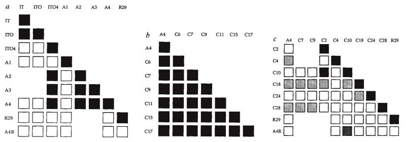
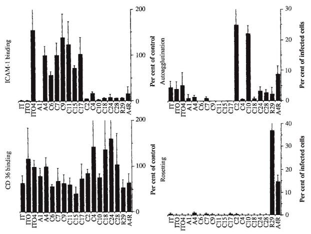
Adhesion of parasitized erythrocytes to post-capillary venular endothelium or uninfected red cells is strongly implicated in the pathogenesis of severe Plasmodium falciparum malaria. Neoantigens at the infected red-cell surface adhere to a variety of host receptors, demonstrate serological diversity in field isolates and may also be a target of the host-protective immune response. Here we use sequential cloning of P. falciparum by micromanipulation to investigate the ability of a parasite to switch antigenic and cytoadherence phenotypes. Our data show that antigens at the parasitized cell surface undergo clonal variation in vitro in the absence of immune pressure at the rate of 2% per generation with concomitant modulations of the adhesive phenotype. A clone has the potential to switch at high frequency to a variety of antigenic and adhesive phenotypes, including a new type of cytoadherence behaviour, 'auto-agglutination' of infected erythrocytes. This rapid appearance of antigenic and functional heterogeneity has important implications for pathogenesis and acquired immunity.


Figures

FIG. 1
FIG. 1
Tree showing derivation of the P. falciparum clones from IT 4/25/5. Clones derived by micromanipulation are shown in the bold boxes. Antigenically indistinguishable clones are boxed together. R29 was cloned from ITOR and A4R derived from A4 by 8 rounds of selection for rosetting. METHODS. Parasites were grown as described, in 95% N2, 1% O2, 4% CO2 using RPMI 1640 culture medium (Gibco) supplemented with 20 mM HEPES, 20 mM glucose, 5 mM glutamine and gentamycin, and 10% pooled serum from blood donors (Blood Transfusion Service, Oxford). During experiments comparing the antigenic phenotype, the cytoadherence characteristics and surface radiolabelling, clones were grown in red blood cells from the same donor and in the same pool of serum. Rosetting parasites were selected for by sedimentation through ‘plasma gel’ (Laboratoire Roger Bellon, Neuilly, France). Parasites were selected for binding to C32 melanoma cells or HUVEC as previously described ,. Cloning was performed using a micromanipulator (Micro Instruments, Oxford), sterile micropipettes of 5 μm internal diameter drawn from capillary tubes and a binocular microscope. Parasitized cells containing single trophozoites were picked from a settled monolayer by applying an aspiration pressure of 1 cm of water, transferred into a Petri dish containing supplemented RPMI 1640 and grown as above. To exclude cross-contamination, a Southern blot of Hinfl-digested DNA from the clones and other laboratory lines was hybridized with the probe pC4. H32 labelled with 32P (Amersham) using a Quickprime labelling kit (Pharmacia). The autoradiograph showed that no cross-contamination had occurred (data not shown).
FIG. 2
FIG. 2
Antigenic analysis of the clones. The antigenic differences between a, IT (clone IT 4/25/5), IT0, IT04, A1-4, R29 and A4R; b, A4 and C clones with the same variant antigenic type as A4; c, A4, C7, C9, variant C clones, R29 and A4. Key: ■, 100 to 80%; square with dark shading, 79 to 15%; lightly shaded squares, 15 to 5%; and □, 5 to 0% similarity between test clones using the mixed agglutination assay. A further four C clones had the same variant type as A4, and the remaining five C clones had a different antigenic type from A4 (data not shown). METHODS. The antigenic phenotype of the derived clones was determined by the mixed agglutination assay (C.I.N. et al., submitted). In brief, the two clones to be tested were labelled independently with either ethidium bromide (200 μg ml−1) or DAPI (4, 6-diamidino-2-methylphenylindole) (20 μg ml−1) for 10 min at 37°C, and washed three times in RPMI 1640. Labelled trophozoites (2.5 × 10⁶) were taken from each of the two cultures and resuspended in 70 μl pooled, heat-inactivated, hyperimmune serum and 140 μl serum-free RPMI 1640. The tube was rotated at 10 r.p.m. at room temperature for 1 h. A drop of the suspension was placed on a microscope slide and sealed with a greased coverslip. Agglutinates were examined using a Leitz epifluorescence microscope with an G365 excitation filter, an FT 395 beam splitter and an LP 420 barrier filter. Agglutinates containing 5 to 10 cells were scored for whether the agglutinate contained all red cells (stained with ethidium bromide), all blue cells (stained with DAPI) or a mixture of red and blue cells. 100 agglutinates were counted in two or more separate experiments for each pair of clones tested. The proportion of mixed-colour agglutinates is a function of the similarity of the antigenic phenotype expressed at the red blood cell surface of the two clones. The distribution of the types of agglutinate can be predicted by the binomial distribution (D.J.R., unpublished observations). If the chance of homologous agglutination is p, then for an agglutinate of size n the chance of the agglutinate being of a single colour is pn−1, and the proportion of mixed-colour agglutinates 1 − pn−1. As equal numbers of parasites from the two clones are used in the test, the ratio of chance of heterologous agglutination/chance of homologous agglutination or 1−p/p is a measure of the degree of similarity between the clones. For example, assuming all agglutinates were of size 5, and the clones were antigenically indistinguishable (p =0.5), the proportion of mixed agglutinates would be 0.94 and the degree of similarity would be 0.5/0.5=1. Similarly, mixed-colour agglutinate proportions of 0.40 and 0.20 in this test represent a degree of similarity between the two clones under test of 0.15 (15%) and 0.05 (5%), respectively.
FIG. 3
FIG. 3
The cytoadherence phenotype of the clones. Adherence of infected red cells to a, purified CD36; b, an ICAM-1–Fc chimaera; c, rosetting of uninfected by infected red cells, and d, auto-agglutination of infected red cells. Binding to CD36 and to the ICAM-1–Fc chimaera is given as the average ± s.d. of two experiments using 4 determinations on each occasion. The percentage of infected erythrocytes rosetting is the mean ± s.d. for 500 infected cells on four occasions, and for auto-agglutination is the mean ± s.d. counting 1,000 infected cells in four determinations. METHODS. CD36 was isolated from human platelets and an ICAM-1–Fc chimaera prepared by transient expression in COS cells and purification on protein G-Sepharose. A solution of CD36 (3 μl; 1 μg ml−1) or ICAM-1–Fc (3 μl; 10 μg ml−1) were adsorbed on to a plastic dish (Falcon no. 1005) in a moist atmosphere at 4 °C overnight. The spots were aspirated and the dish washed five times with binding medium (RPMI, HEPES 20 mM, pH 6.9) before blocking with 0.5% BSA/RPMI 1640 for 4 h at 37 °C and washing 5 times in binding medium. A suspension of trophozoites (5 ml) in binding medium at 1% haematocrit and between 1 and 2% parasitaemia was placed in the dish, incubated at 37 °C, resuspending the red cells every 10 min. This was repeated five times. Unbound cells were removed from the dish by careful washing. The adherent cells were fixed with 1% glutaraldehyde, stained with Giemsa and the number of cells per high power field counted. The counts were normalized to the binding of A4 clone for both CD36 and ICAM-1 binding in each experiment. Rosetting was measured by counting the number of trophozites rosetting two or more uninfected red cells immediately after removing a sample from the culture flask. The percentage of trophozites agglutinating with other infected cells (that is, auto-agglutinating) was counted after 200 μl culture at 10% haematocrit was mixed at 10 r.p.m. for 1 h.
FIG. 4
FIG. 4
The relative a, antigenic and b, adhesive phenotypes of the parental clone A4, progeny clone C28 and line C28-1 derived from C28 by selection once on ICAM-1. Key: ■, 100 to 80%; shaded square, 79 to 25%; and □, 15 to 0% similarity between clones using the mixed agglutination assay. Binding to CD36 and to the ICAM-1–Fc chimaera are given as the average ± s.d. of four determinations. Results of a representative experiment are shown. METHODS. ICAM-1–Fc chimaera (50 μg ml−1 in PBS/gentamycin) was adsorbed on to a plastic dish (Falcon no. 1005) in a moist atmosphere at 37 °C for 2 h. The solution was removed and the dish blocked with 2% BSA/PBS/gentamycin for 2 h at 37 °C and finally wahed three times in binding medium. A suspension of late trophozoites (1.25 ml) purified by gelatin flotation was allowed to settle on the dish for 1 h before the unbound parasites were washed off. Erythrocytes (50 μl) were added to the bound cells in culture medium, and the dish placed in a gas jar at 37 °C overnight. Red cells were collected the next day and cultured as described (Fig. 1). The mixed agglutination and cytoadherence assays were done as described.

Comment in

Similar articles

Cited by

References

    1. MacPherson GG, Warrell MJ, White NJ, Looareesuran S, Warrell DA. Am. J. Path. 1985;119:385–401. - PMC - PubMed
    1. David PH, Handunetti SM, Leech JH, Gamage P, Mendis KM. Am. J. trop. Med. Hyg. 1988;38:289–297. - PubMed
    1. Udomsangpetch R, et al. J. exp. Med. 1989;169:1835–1840. - PMC - PubMed
    1. Carlson J, et al. Lancet. 1990;336:1457–1460. - PubMed
    1. Barnwell JW, et al. J. clin. Invest. 1989;84:765–772. - PMC - PubMed

Publication types

Substances