

Case Reports
. 2004 May;14(2):109-13; discussion 113-4.
doi: 10.1055/s-2004-828705.
Malignant peripheral nerve sheath tumor of the orbit: case report and literature review
Affiliations
- PMID: 16145592
- PMCID: PMC1151679
- DOI: 10.1055/s-2004-828705
Item in Clipboard
Case Reports
Malignant peripheral nerve sheath tumor of the orbit: case report and literature review
Skull Base.
2004 May.
Abstract
A 68-year-old woman with progressive visual loss and exophthalmos in her right eye had been operated on for a mass in her right calf 3 years earlier. Imaging showed a huge mass invading the orbital structures and temporal pole. The presumptive diagnosis was a malignant orbital tumor. The tumor was resected totally and eroded tissues such as the lateral rectus muscle and dural compartments were repaired. The histological diagnosis was a malignant peripheral nerve sheath tumor (MPNST). The patient recovered uneventfully and was discharged 8 days after surgery. Two years later she died from a liver tumor. Few MPNSTs involving the orbit have been reported.
Figures
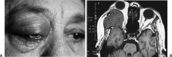
(A) Prominent exophthalmos, chemosis, and flashing in the palpebras in the patient's right eye. (B) T1-weighted MRI showing the lesion originating from the lateral part of the orbit.
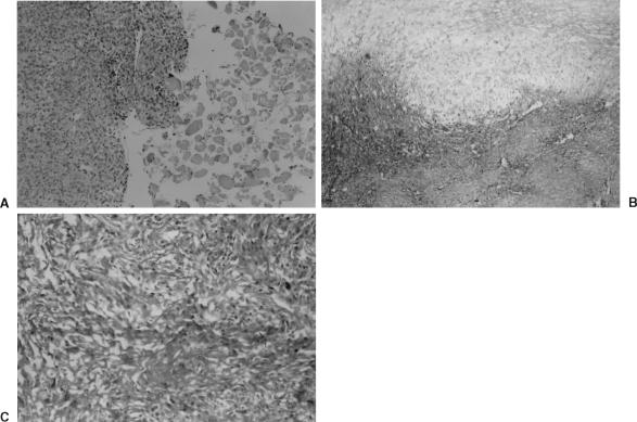
(A) Malignant tumoral cells and infiltrated tissue of the lateral rectus muscle (hematoxylin and eosin (H&E), × 400). (B) Mixoid changes are visible (H&E, × 100). (C) Rounded and ovoid cells include dense vascular and high mitotic activity (H&E, × 100).
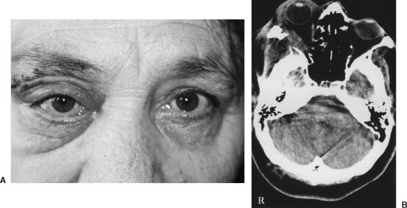
(A) Postoperatively, the patient had no obvious cosmetic problems and deviation of her eye resolved. (B) Postoperative computed tomography showed no residual mass in the orbit 6 months after surgery. The repaired lateral rectus muscle appears normal.
References
-
- D'Agostino A N, Soule E H, Miller R H. Sarcomas of the peripheral nerves and somatic soft tissue associated with multiple neurofibromatosis (von Recklinghausen's disease) Cancer. 1963;16:1015–1027. - PubMed
-
- Grnja V, Allen W E, Osborn D J, Kier E L. Sacral neurofibrosarcoma: an angiographic evaluation. Case report. J Neurosurg. 1974;40:767–771. - PubMed
-
- Wanebo J, Malik J, Vanden Berg S, Wanebo H, Driesen N, Persing J. Malignant peripheral nerve sheath tumors. Cancer. 1993;71:1247–1253. - PubMed
-
- Wick M, Swanson P, Scheithauer B, Manivel J. Malignant peripheral nerve sheath tumor. Am J Clin Pathol. 1987;87:425–433. - PubMed
-
- Kchouk M, Rabet A M, Ghedas K, et al. Extensive malignant schwannoma of the sciatic nerve. Contribution of imaging techniques [in French] J Radiol. 1993;74:641–644. - PubMed
