Review
. 2006 Jan 1:11:81-8.
doi: 10.2741/1781.

Changes in laminin isoforms associated with brain tumor invasion and angiogenesis

Affiliations
Review

Changes in laminin isoforms associated with brain tumor invasion and angiogenesis

Julia Y Ljubimova et al. Front Biosci. .

Abstract

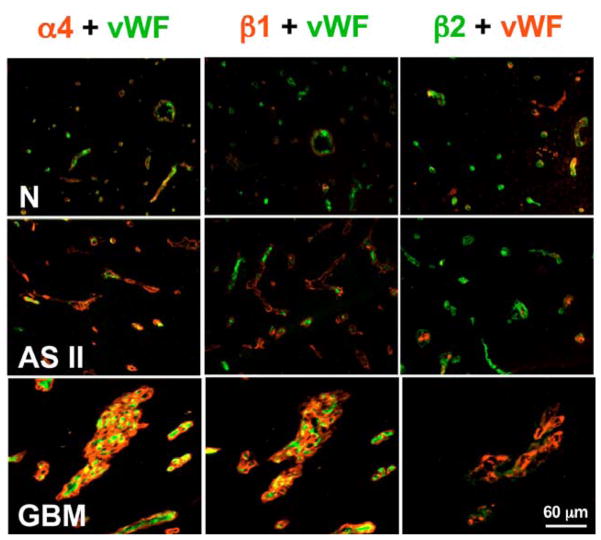
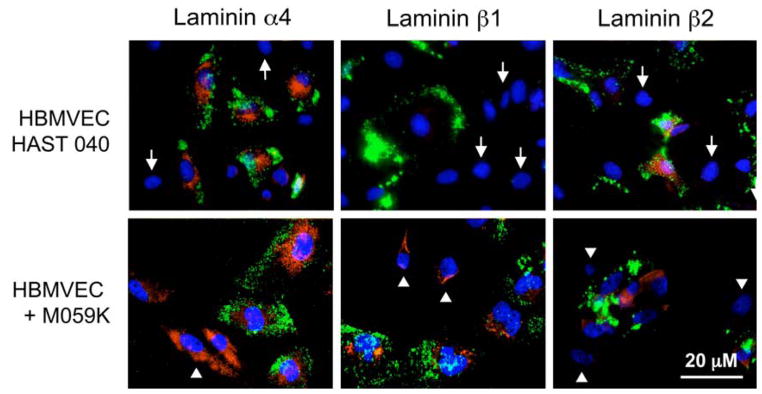
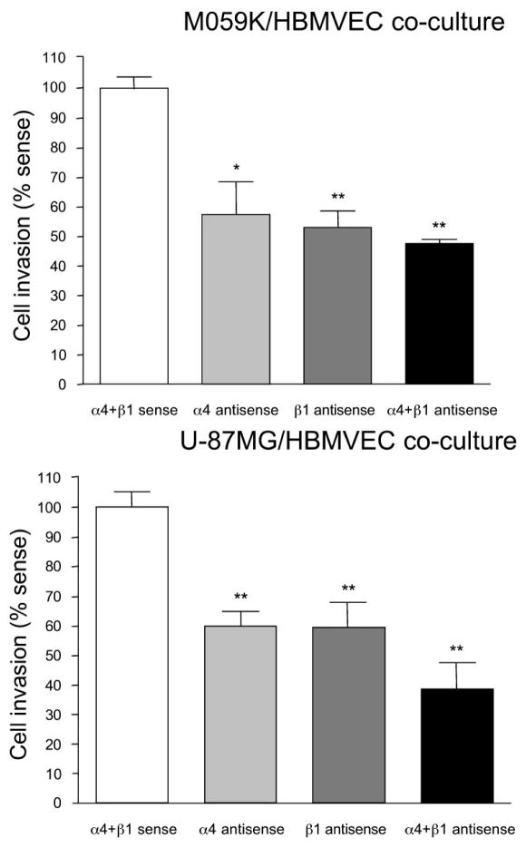
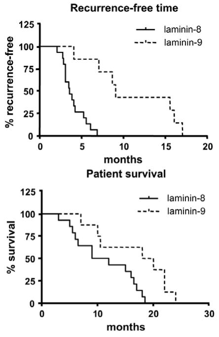
Laminins are the major constituents of blood vessel basement membranes (BMs). Each laminin is a trimer consisting of three assembled polypeptide chains, alpha, beta and gamma. More than 15 laminin isoforms are known to date and the expression of specific isoforms may change in certain pathological conditions. Here we show that during progression of glial tumors laminin-9 (alpha4beta2gamma1) is switched to laminin-8 (alpha4beta1gamma1), which is dramatically increased in glial brain tumors. Laminin-8 overproduction by glial tumor cells facilitates spread of glioma. Brain tumors with laminin-8 overexpression recur faster after standard treatment and patients have shorter survival time. Laminin-8 may be thus used as a predictor of tumor recurrence, patient survival and as a potential molecular target for glioma therapy.


Figures

Figure 1
Figure 1
Double immunofluorescent staining of brain tissues for laminin-8 (α4β1γ1) and laminin-9 (α4β2γ1) chains, and for von Willebrand factor (vWF) specific for endothelial cells. N, Normal brain, where microvessels are positive for vWF, and α4 and β1 laminin chains are barely visible. At the same time, β2 laminin chain is prominent in vessel walls positive for vWF. This pattern is compatible with small amounts of laminin-9. AS II, Astrocytoma grade II with stronger staining for laminin α4 chain in brain microvessels. Expression of laminin β1 chain is higher than in normal brain and β2 is still strong. This pattern is compatible with predominance of laminin-9.GBM, glioblastoma multiforme with very bright staining of α4 and β1 laminin chains but very weak β2 chain in brain vessels. This pattern is compatible with predominance of laminin-8. Reproduced with permission from Cancer 101, 604–612 (2004)
Figure 2
Figure 2
Laminin α4, β1, and β2 chain staining of co-cultures. Live co-cultures were exposed to Ac-LDL (green color, to reveal endothelial cells) and then fixed and simultaneously stained for select laminin chains (red color) and nuclei (DAPI, blue color). In endothelial-normal astrocyte co-cultures (HBMVEC+HAST040) α4 and β2 chains are expressed in Ac-LDL-positive endothelial cells only but not in Ac-LDL-negative astrocytes (arrows). β1 chain is largely absent. In endothelial-glioma co-cultures (HBMVEC+M059K), α4 chain is expressed by both cell types and β2 chain, only by endothelial cells. Importantly, β1 chain is now expressed not only by Ac-LDL-negative glioma cells (arrowheads) but also by Ac-LDL-positive endothelial cells. Reproduced with permission from: Mol Cancer Ther 2, 985–994 (2003)
Figure 3
Figure 3
Measurement of invasion in co-cultures after antisense treatment. The Matrigel invasion assay was carried out as described in Materials and Methods. Note significant decrease in the fraction of cells that invaded through Matrigel in antisense-treated cultures. A more pronounced effect is seen with a combination of antisense oligonucleotides. Similar results were obtained with M059K and U-87MG glioma cell lines. *, p<0.04; **, p<0.001 by ANOVA. Invasion in sense-treated cultures was taken as 100%. Reproduced with permission from: Mol Cancer Ther 2, 985–994 (2003)
Figure 4
Figure 4
Laminin-8 associates with decreased time of recurrent tumor development and decreased survival of GBM patients compared to laminin-9. Statistical analysis was done using Kaplan-Meier test. p=0.0002 for tumor recurrence, and p<0.015 for patient survival. Reproduced with permission from Cancer 101, 604–612 (2004)

References

    1. Shapiro WR, Shapiro JR. Biology and treatment of malignant glioma. Oncology. 1998;12:233–240. - PubMed
    1. Gonzales MF. Classification and pathogenesis of brain tumors. In: Kaye AH, Laws ER Jr, editors. Brain tumors. Churchill Livingstone; Edinburgh: 1997.
    1. Ljubimova JY, Khazenzon NM, Chen Z, Neyman YI, Turner L, Riedinger MS, Black KL. Gene array analysis of differentially expressed genes in human glial tumors. Int J Oncol. 2001;18:287–295. - PubMed
    1. Ljubimova JY, Lakhter AJ, Loksh A, Yong WH, Riedinger MS, Miner JH, Sorokin ML, Ljubimov AV, Black KL. Overexpression of α4 chain-containing laminins in human glial tumors identified by gene microarray analysis. Cancer Res. 2001;61:5601–5610. - PubMed
    1. Sehgal A. Molecular changes during the genesis of human gliomas. Semin Surg Oncol. 1998;14:3–12. - PubMed

MeSH terms

LinkOut - more resources