Comparative Study
. 2005 Sep 5;202(5):687-96.
doi: 10.1084/jem.20051035.

Adhesive mechanisms governing interferon-producing cell recruitment into lymph nodes

Affiliations
Comparative Study

Adhesive mechanisms governing interferon-producing cell recruitment into lymph nodes

Thomas G Diacovo et al. J Exp Med. .

Abstract

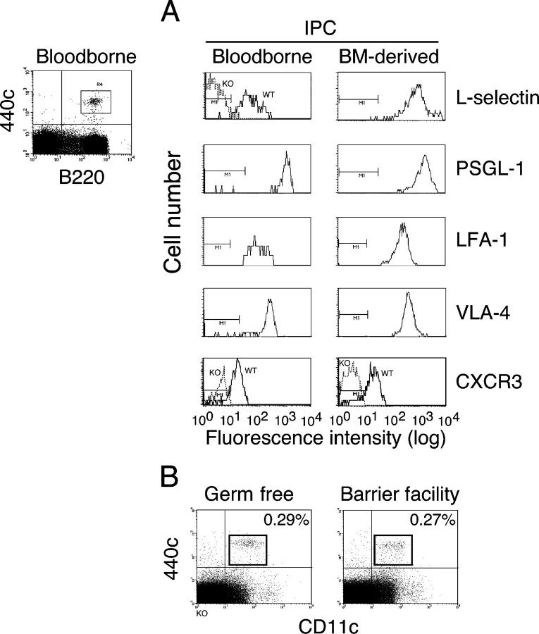
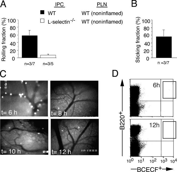
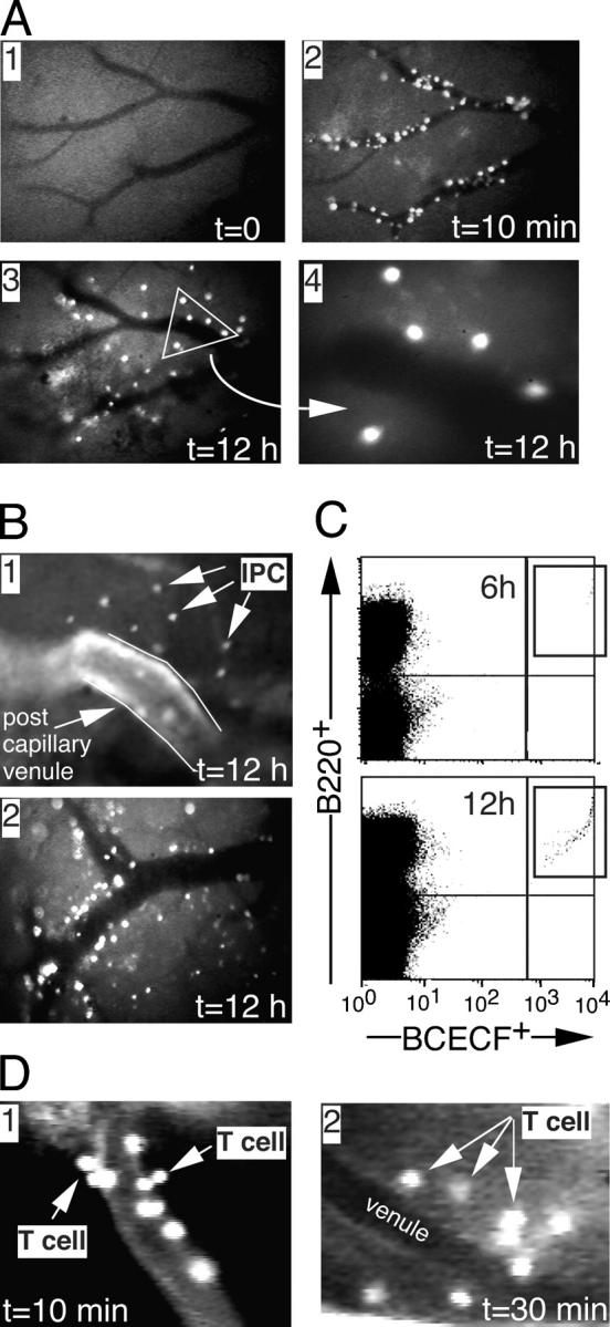
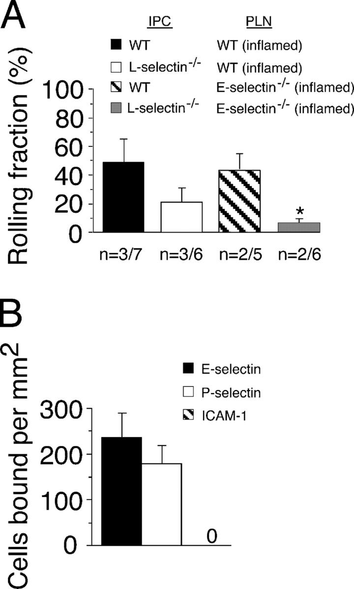
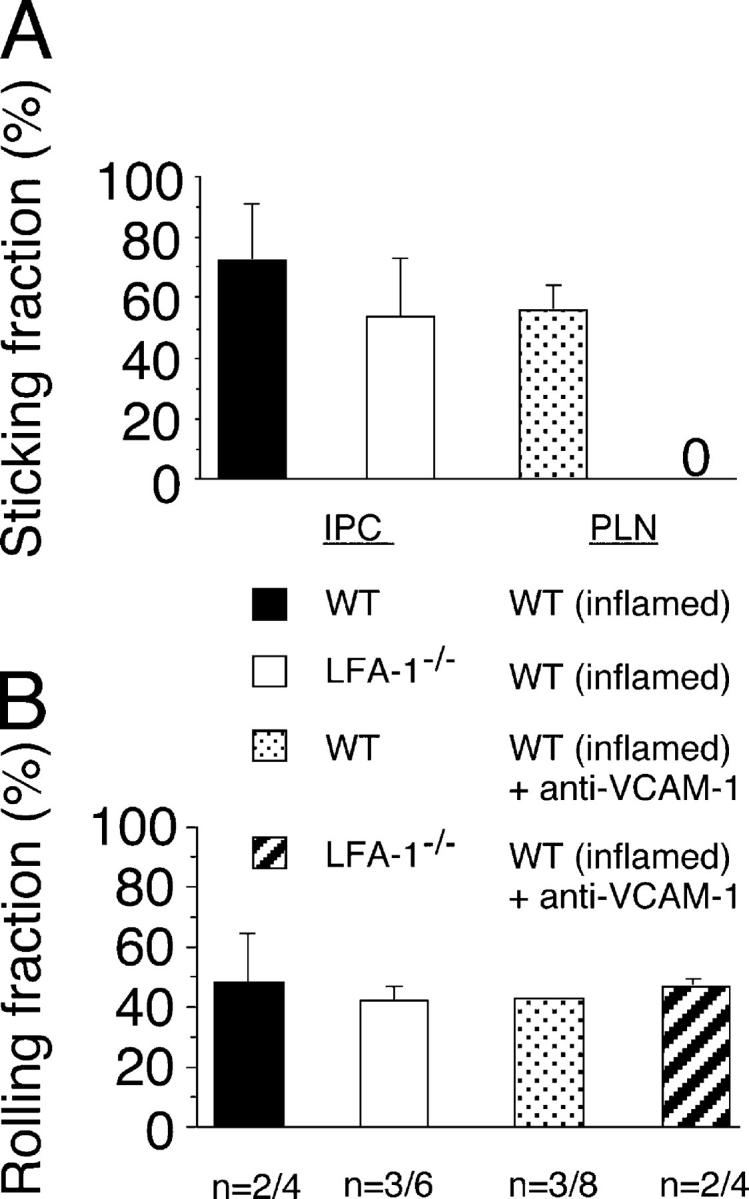
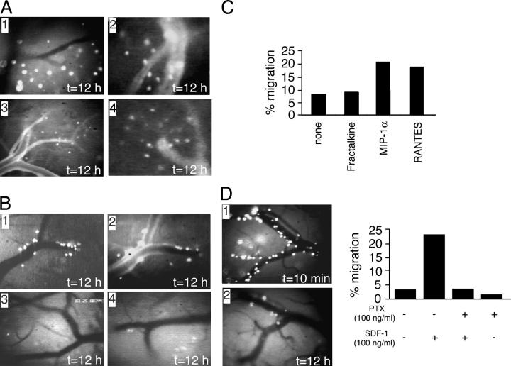
Natural interferon-producing cells (IPCs) are found in peripheral lymph nodes (PLNs), where they support NK cell, T cell, and B cell responses to pathogens. However, their route of entry and the adhesive mechanisms used to gain access to PLNs remain poorly defined. We report that IPCs can enter PLNs via a hematogenous route, which involves a multistep adhesive process, and that transmigration is enhanced by inflammation. Results indicate that L-selectin on IPCs is required for efficient attachment and rolling on high endothelial venules in vivo in both nonstimulated and inflamed PLNs. IPCs, however, also possess functional ligands for E-selectin that contribute to this process only in the latter case. In conjunction with selectin-mediated adhesion, both beta(1)- and beta(2)-integrins participate in IPC attachment to the inflamed vessel wall, whereas chemotaxis relies in part on the chemokine receptor CCR5. Identification of the adhesive machinery required for IPC trafficking into PLNs may provide opportunities to regulate immune responses reliant on the activity of these cells.


Figures

Figure 1.
Figure 1.
Surface expression of homing molecules on bloodborne- and BM-derived murine IPCs. IPCs were identified in either heparinized whole blood or BM cultured with Flt3 ligand by gating on B220+/440c+ cells. Note that these cells were also CD11c+ and CD11blow/− (not depicted). Both bloodborne and culture-derived IPCs express L-selectin, PSGL-1, LFA-1, VLA-4, and CXCR3. KO controls are also shown for L-selectin and CXCR3 for comparison. (B) Representative dot plot demonstrating the presence of IPCs in PLNs of germ-free mice as compared with those housed solely in a barrier facility. The percentage of cells in each gate is indicated.
Figure 2.
Figure 2.
IPC roll and stick but minimally transmigrate across HEVs in noninflamed PLNs. BCECF-labeled IPCs, generated from the BM of WT or L-selectin−/− mice, were injected into the microcirculation of a noninflamed PLN, and the (A) percent rolling fraction and (B) percent sticking fraction were determined by fluorescence microscopy (20×) in identical fields of view. n = number of mice per venules analyzed. Data are shown as mean ± SE. (C) Representative micrographs of HEVs in PLNs at 6, 8, 10, or 12 h after injection of labeled IPCs (10×). The skin overlying the subiliac LN was initially left intact until the designated time of viewing. Data are representative of three individual experiments. (D) Representative dot plot to evaluate the accumulation of BCECF-labeled WT IPCs in two noninflamed subiliac PLNs at 6 or 12 h after injection, as determined by flow cytometry (2 vs. 1 BCECF-labeled cells/105 total events, respectively).
Figure 3.
Figure 3.
IPC transmigration into PLNs is enhanced by inflammation. Representative micrographs depicting (A) HEVs alone (panel 1, 10×), HEVs 10 min after injection of BCECF-labeled WT IPCs (panel 2, 10×), HEVs 12 h after injection of labeled cells (panel 3, 10×), and an enlarged section of PLNs (panel 4, 20×). (B) Representative micrographs of an inflamed PLN after i.v. injection of 150 kD FITC–dextran and 12h after injection of BCECF-labeled WT IPCs (panel 1, 40×). Lines demarcate lateral borders of the lumenal compartment. A micrograph demonstrating IPC transmigration at 12 h without prior surgical intervention is shown for comparison (panel 2). PLN inflammation was induced by injection of Mtb as described in Materials and methods. (C) Representative dot blot to evaluate the accumulation of BCECF-labeled WT IPCs in inflamed subiliac PLNs at 6 or 12 h after injection, as determined by flow cytometry (2 vs. 13 BCECF-labeled cells/105 total events, respectively). (D) Representative micrographs of an inflamed PLN after i.v. administration of 150 kD FITC–dextran at 10 and 30 min after injection of BCECF-labeled T cells (panels 1 and 2, respectively; 40×).
Figure 4.
Figure 4.
L- and E-selectin are required for IPC attachment and rolling on HEVs of inflamed PLNs. BCECF-labeled IPCs generated from the BM of WT or L-selectin−/− mice were injected through the femoral artery of WT or E-selectin−/− animals, and the rolling fraction was quantified as they passed through HEVs in inflamed PLNs. Data are shown as mean ± SE. *, P < 0.05; n = number of mice per venules analyzed. (B) Attachment of WT IPCs to surface-immobilized E- or P-selectin Ig chimeras at a wall shear rate of 200 s-1. Adhesion to the β2-integrin ligand ICAM-1–Ig chimera is shown as a negative control. Results represent the mean ± SE for three experiments performed in duplicate.
Figure 5.
Figure 5.
β 1- and β 2-integrins on IPCs participate in firm adhesion. The percent sticking fraction of WT or LFA-1−/− IPCs in HEVs of inflamed PLNs in the absence or presence of the VCAM-1 function blocking F(ab′)2 M/K-2.7 was evaluated during their initial passage through the microcirculation. Data are shown as mean ± SE. n = number of mice per venules analyzed. (B) The percent rolling fraction of IPCs under the identical conditions.
Figure 6.
Figure 6.
IPC transmigration across HEVs in inflamed PLNs is CCR5-dependent. (A) Representative micrographs of HEVs in inflamed PLNs 12 h after injection of CXCR3−/− IPCs in the absence of (panel 1, 10×) or after i.v. administration of FITC–dextran to delineate the vascular compartment (panel 2, 10×; panels 3 and 4, 20×). All panels are from individual animals (n = 4). (B) Representative micrographs of HEVs in inflamed PLNs 12 h after injection of CCR5−/− IPCs in the absence of (panels 1, 3, and 4; 10×) or after i.v. administration of (panel 2, 10×) FITC–dextran to delineate the vascular compartment. All panels are from individual animals (n = 3). (C) Migration of BM-derived IPCs across bare transwell filters in response to fractalkine, MIP-1α, or RANTES. (D) Representative micrographs depicting the same HEV either 10 min (panel 1, 10×) or 12 h (panel 2, 10×) after the injection of BCECF-labeled WT IPCs pretreated with 100 ng/ml PTX. The effect of 100 ng/ml PTX on SDF-1–induced chemotaxis in an in vitro transwell assay system is shown for comparison.

References

    1. Colonna, M., G. Trinchieri, and Y.J. Liu. 2004. Plasmacytoid dendritic cells in immunity. Nat. Immunol. 5:1219–1226. - PubMed
    1. Liu, Y.J. 2005. IPC: professional type 1 interferon-producing cells and plasmacytoid dendritic cell precursors. Annu. Rev. Immunol. 23:275–306. - PubMed
    1. Iwasaki, A., and R. Medzhitov. 2004. Toll-like receptor control of the adaptive immune responses. Nat. Immunol. 5:987–995. - PubMed
    1. Siegal, F.P., N. Kadowaki, M. Shodell, P.A. Fitzgerald-Bocarsly, K. Shah, S. Ho, S. Antonenko, and Y.J. Liu. 1999. The nature of the principal type 1 interferon-producing cells in human blood. Science. 284:1835–1837. - PubMed
    1. Cella, M., F. Facchetti, A. Lanzavecchia, and M. Colonna. 2000. Plasmacytoid dendritic cells activated by influenza virus and CD40L drive a potent TH1 polarization. Nat. Immunol. 1:305–310. - PubMed

Publication types

LinkOut - more resources