. 2005 Sep 7;25(36):8115-21.
doi: 10.1523/JNEUROSCI.1143-05.2005.

Cdh1/Hct1-APC is essential for the survival of postmitotic neurons

Affiliations

Cdh1/Hct1-APC is essential for the survival of postmitotic neurons

Angeles Almeida et al. J Neurosci. .

Abstract

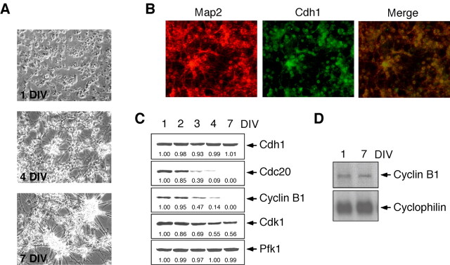
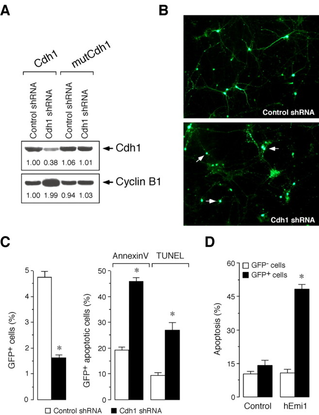
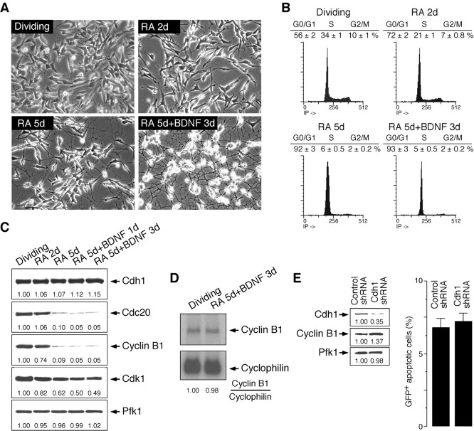
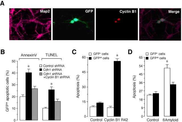
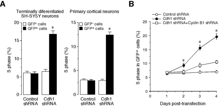
Cell division at the end of mitosis and G1 is controlled by Cdh1/Hct1, an activator of the E3-ubiquitin ligase anaphase-promoting complex (APC) that promotes the ubiquitylation and degradation of mitotic cyclins and other substrates. Cdh1-APC is active in postmitotic neurons, where it regulates axonal growth and patterning in the developing brain. However, it remains unknown whether Cdh1-APC is involved in preventing cell-cycle progression in terminally differentiated neurons. To address this issue, we used the small hairpin RNA strategy to deplete Cdh1 in postmitotic neurons. We observed that Cdh1 silencing rapidly triggered apoptotic neuronal death. To investigate the underlying mechanism, we focused on cyclin B1, a major Cdh1-APC substrate. Our results demonstrate that Cdh1 is required to prevent the accumulation of cyclin B1 in terminally differentiated neurons. Moreover, by keeping cyclin B1 low, Cdh1 prevented these neurons from entering an aberrant S phase that led to apoptotic cell death. These results provide an explanation for the mechanism of cyclin B1 reactivation that occurs in the brain of patients suffering from neurodegenerative diseases, such as Alzheimer's disease.


Figures

Figure 1.
Figure 1.
Cdh1, but not Cdc20, is present in postmitotic neurons. A, Phase-contrast microscopy of E16 rat cortical neurons along in vitro differentiation at 1, 4, and 7 DIV showing neurite processes as from 4 DIV. B, Identification of 7 DIV neuronal cultures by immunocytochemistry showing that all Map2-positive cells expressed Cdh1. C, Western blot analyses confirmed Cdh1 expression throughout terminal differentiation of cortical neurons in primary culture (from 1 to 7 DIV), whereas Cdc20 and cyclin B1 decreased progressively to undetectable levels after 7 DIV, and Cdk1 was still present in neurons after 7 DIV. D, Cyclin B1 mRNA was unchanged from 1 to 7 DIV. Pfk1 (6-phosphofructo-1-kinase) and cyclophilin were used as loading controls for protein and RNA, respectively.
Figure 2.
Figure 2.
Cdh1 is essential for the survival of postmitotic neurons. A, Cdh1 shRNA is specific, as assessed by expression of target gene refractory to the RNA interfering sequence. Thus, transfection of 293T cells with control shRNA (pSuper.GFP.luciferase) did not alter the protein levels of expressed Cdh1, regardless of the Cdh1 cDNA coexpressed (wild type or mutant). However, transfection of cells with Cdh1 shRNA (pSuper.GFP.Cdh1) decreased Cdh1 protein levels expressed from wild-type (Cdh1) but not from mutant (mutCdh1) Cdh1. Moreover, endogenous cyclin B1 protein levels increased in those cells having lower Cdh1 protein levels. B, Fluorescence images show neurite disintegration and cell body condensation in cortical primary neurons transfected with Cdh1 shRNA compared with the control shRNA. C, Transfection of cortical primary neurons with pSuper.GFP.Cdh1 (Cdh1 shRNA) decreased the number of GFP+ cells (left) and increased apoptotic cells within the GFP+ subpopulation, as assessed both by annexin-V and TUNEL (right); neurons transfected with pSuper.GFP.luciferase were used as controls (control shRNA). D, Transfection of primary neurons with APC inhibitor hEmi1 increased the number of apoptotic cells within the GFP+ subpopulation but not within the GFP subpopulation compared with control cells expressing a nonfunctional truncated form of hEmi1. *p < 0.05 versus the corresponding control shRNA.
Figure 3.
Figure 3.
Characterization of SH-SY5Y neuroblastoma cells subjected to terminal differentiation and effect of Cdh1 depletion in the dividing SH-SY5Y cells. A, Phase-contrast microscopy of differentiating SH-SY5Y cells, showing the morphology of the postmitotic neuronal phenotype. B, Cell-cycle distribution of SH-SY5Y cells along the differentiation process, showing G0/G1 arrest after full differentiation. C, Western blot analysis revealed that Cdh1 levels were maintained and Cdk1 levels were present from the dividing to the terminally differentiated stages, whereas cyclin B1 and Cdc20 levels decreased. D, Cyclin B1 mRNA was maintained in terminally differentiated SH-SY5Y cells. E, Depletion of Cdh1 in dividing SH-SY5Y cells triggered cyclin B1 protein stabilization (left) but did not affect cell survival (right).
Figure 4.
Figure 4.
Cdh1 promotes survival of terminally differentiated SH-SY5Y cells by preventing cyclin B1 accumulation. A, Representative confocal microscopy image (6 cells in this field) of terminally differentiated SH-SY5Y cells 3 d after Cdh1 shRNA treatment, indicating that only efficiently transfected cells (identified as GFP+ cells; 2 cells in this field) displayed cyclin B1 immunoreactivity (identified as red fluorescent cells). In contrast, nontransfected cells (identified as GFP cells) showed no cyclin B1 immunoreactivity. The same pattern was observed in all fields examined. B, Cyclin B1 shRNA is specific, as assessed by expression of target gene refractory to the RNA interfering sequence. Thus, transfection of 293T cells with control shRNA (pSuper.GFP.luciferase) did not alter the protein levels of expressed cyclin B1, regardless of the cyclin B1 cDNA coexpressed (wild type or mutant). However, transfection of cells with cyclin B1 shRNA (pSuper.GFP.cyclin B1) decreased cyclin B1 protein levels expressed from wild-type (cyclin B1) but not from mutant (mutCyclin B1) cyclin B1. Endogenous Cdh1 protein levels were unaffected by cyclin B1 shRNA. C, Cyclin B1 shRNA prevented the increase in cyclin B1 triggered by Cdh1 shRNA in 293T cells. D, Cdh1 silencing in terminally differentiated SH-SY5Y cells triggered an increase in the proportion of GFP+ apoptotic cells. This effect was prevented by cotransfection with cyclin B1 shRNA. *p < 0.05 versus corresponding control shRNA. Control shRNA, pSuper.GFP.luciferase plus pSuper; Cdh1 shRNA, pSuper.GFP.Cdh1 plus pSuper; cyclin B1 shRNA, pSuper.GFP.Cdh1 plus pSuper.cyclin B1.
Figure 5.
Figure 5.
Cdh1 promotes survival of primary cortical neurons by preventing cyclin B1 accumulation. A, Representative fluorescence microscopy image of 7 DIV neurons (identified as Map2-positive cells) 3 d after Cdh1 shRNA treatment, indicating that only efficiently transfected cells (identified as GFP+ cells; 2 cells in this field) displayed cyclin B1 immunoreactivity (identified as red fluorescent cells). The localization of cyclin B1 was displayed in a condensed manner, suggesting nuclear localization. In contrast, nontransfected cells (identified as GFP cells) showed no cyclin B1 immunoreactivity. The same pattern was observed in all fields examined. B, Cdh1 silencing in primary cortical neurons triggered an increase in the proportion of GFP+ apoptotic cells, as assessed both by annexin-V and TUNEL. This effect was prevented by cotransfection with cyclin B1 shRNA. Control shRNA, pSuper.GFP.luciferase plus pSuper; Cdh1 shRNA, pSuper.GFP.Cdh1 plus pSuper; cyclin B1 shRNA, pSuper.GFP.Cdh1 plus pSuper.cyclin B1. C, Overexpression of cyclin B1 by transfection of cortical primary neurons with a plasmid construction encoding a nondegradable form of cyclin B1 fused to GFP (cyclin B1-R42) increased apoptotic cells within the GFP+ subpopulation but not within the GFP subpopulation compared with control cells. D, Overexpression of Cdh1 in cortical primary neurons did not affect neuronal survival but significantly prevented apoptotic neuronal death triggered by β-amyloid. *p < 0.05 versus corresponding control shRNA.
Figure 6.
Figure 6.
Depletion of Cdh1 promotes cyclin B1-mediated re-entry of postmitotic neurons into S phase. A, Transfection of terminally differentiated SH-SY5Y or cortical neurons with Cdh1 shRNA increased the proportion of cells in S phase, as assessed by BrdU incorporation. B, Cyclin B1 shRNA counteracted the increase in the proportion of postmitotic SH-SY5Y cells entering S phase triggered by Cdh1 silencing. *p < 0.05 versus corresponding control. Control shRNA, pSuper.GFP.luciferase plus pSuper; Cdh1 shRNA, pSuper.GFP.Cdh1 plus pSuper; cyclin B1 shRNA, pSuper.GFP.Cdh1 plus pSuper.cyclin B1.

References

    1. Almeida A, Moncada S, Bolaños JP (2004) Nitric oxide switches on glycolysis through the AMP protein kinase and 6-phosphofructo-2-kinase pathway. Nat Cell Biol 6: 45–51. - PubMed
    1. Bashir T, Dorrello NV, Amador V, Guardavaccaro D, Pagano M (2004) Control of the SCF(Skp2-Cks1) ubiquitin ligase by the APC/C(Cdh1) ubiquitin ligase. Nature 428: 190–193. - PubMed
    1. Becker EBE, Bonni A (2004) Cell cycle regulation of neuronal apoptosis in development and disease. Prog Neurobiol 72: 1–25. - PubMed
    1. Blanco MA, Sanchez-Diaz A, de Prada JM, Moreno S (2000) APC(ste9/srw1) promotes degradation of mitotic cyclins in G(1) and is inhibited by cdc2 phosphorylation. EMBO J 19: 3945–3955. - PMC - PubMed
    1. Brummelkamp TR, Bernards R, Agami R (2002) A system for stable expression of short interfering RNAs in mammalian cells. Science 296: 550–553. - PubMed

Publication types

MeSH terms

LinkOut - more resources