. 2005 Sep 7;25(36):8229-39.
doi: 10.1523/JNEUROSCI.1469-05.2005.

Recurrent mossy fibers establish aberrant kainate receptor-operated synapses on granule cells from epileptic rats

Affiliations

Recurrent mossy fibers establish aberrant kainate receptor-operated synapses on granule cells from epileptic rats

Jérôme Epsztein et al. J Neurosci. .

Abstract

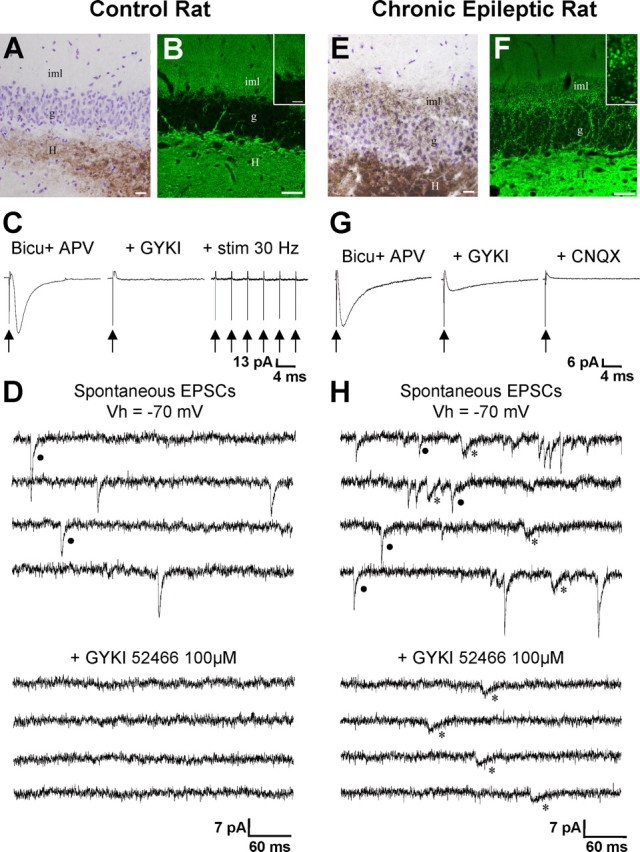
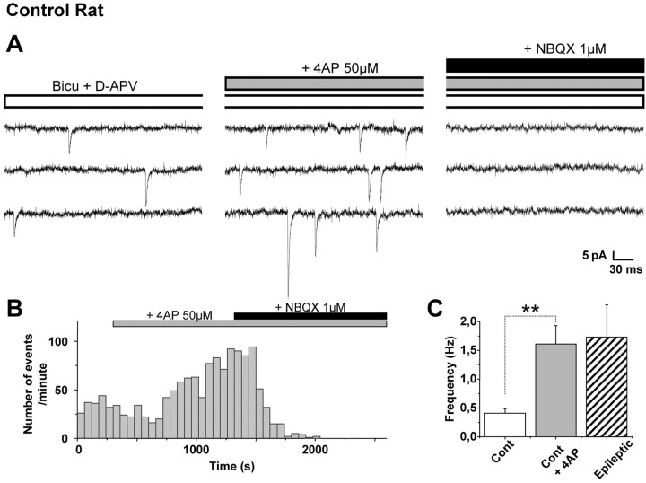
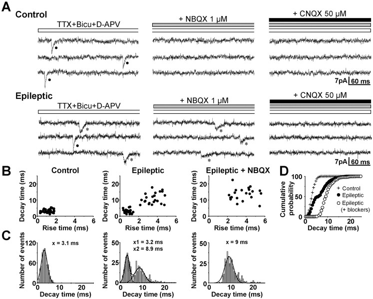
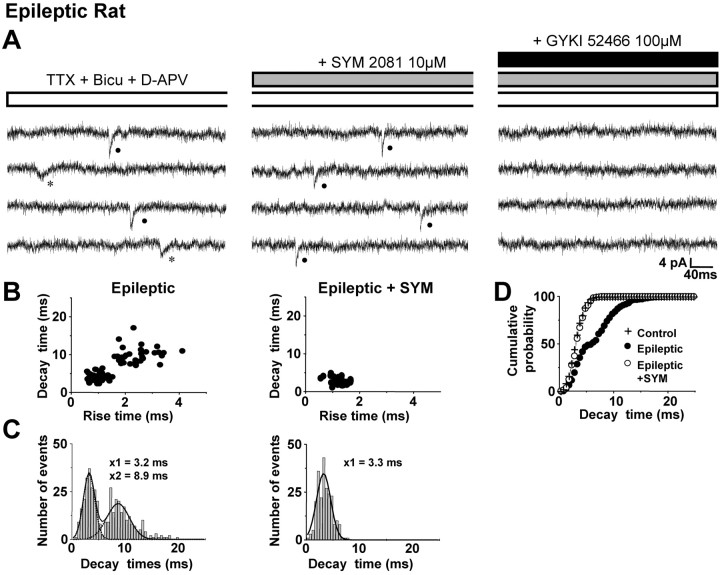
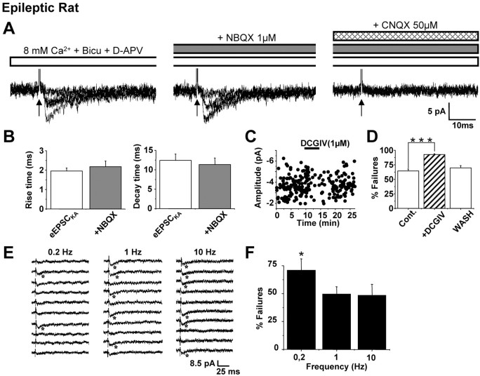
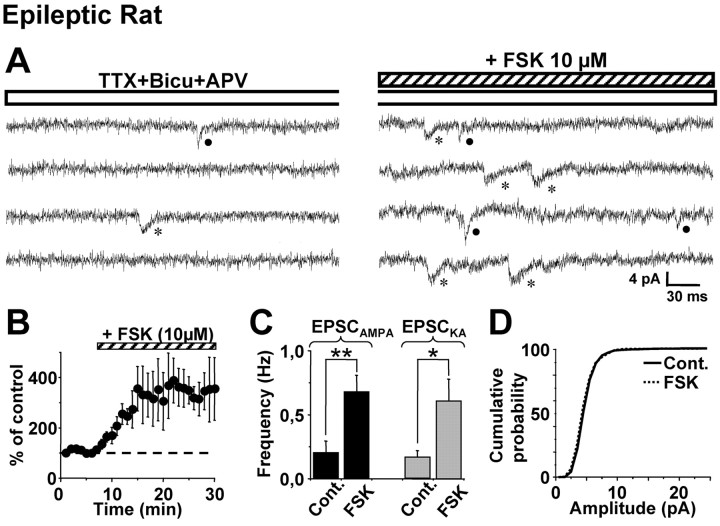
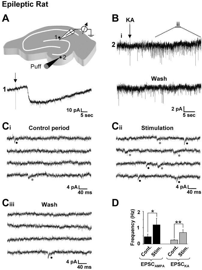
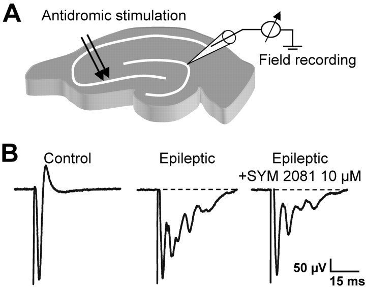
Glutamatergic mossy fibers of the hippocampus sprout in temporal lobe epilepsy and establish aberrant synapses on granule cells from which they originate. There is currently no evidence for the activation of kainate receptors (KARs) at recurrent mossy fiber synapses in epileptic animals, despite their important role at control mossy fiber synapses. We report that KARs are involved in ongoing glutamatergic transmission in granule cells from chronic epileptic but not control animals. KARs provide a substantial component of glutamatergic activity, because they support half of the non-NMDA receptor-mediated excitatory drive in these cells. KAR-mediated EPSC(KA)s are selectively generated by recurrent mossy fiber inputs and have a slower kinetics than EPSC(AMPA). Therefore, in addition to axonal rewiring, sprouting of mossy fibers induces a shift in the nature of glutamatergic transmission in granule cells that may contribute to the physiopathology of the dentate gyrus in epileptic animals.


Figures

Figure 1.
Figure 1.
Mossy fiber sprouting and EPSCKA in granule cells from epileptic rats. A, E, Timm staining (dark brown; counterstained with cresyl violet) of mossy fibers of control (A) and epileptic (E) rat sections after electrophysiological recordings. In chronic epileptic rats, mossy fiber collaterals invade the granule cell layer (g) and the inner third of the molecular layer (iml). Scale bars, 20μm. B, F, VGLUT1 immunostaining of sections from control (B) and epileptic (F) rats. In epileptic rats, the staining dramatically increases in the inner molecular layer (iml) and granule cell layer, in good correlation with the sprouting of mossy fibers. In the inner molecular layer, the type of staining is modified, as large spots (characteristic of the staining of large mossy fiber synaptic boutons) have replaced the fine speckled staining observed in the control (enlarged in the insets). Scale bars, 50μm; insets, 10μm. C, G, Averages of 30 individual EPSCs to stimulations (0.033 Hz) in the inner one-third of the molecular layer of the dentate gyrus recorded in the presence of 10μm bicuculline (Bicu) and 40μm d-APV (APV). In control granule cells (C), the EPSC evoked in control conditions (left) was completely blocked by AMPA receptor antagonist GYKI 52466(GYKI; 100 μm; middle), even when repetitive stimulations were applied (10 stimuli at 30 Hz; stim 30 Hz; right). In dentate GCs from epileptic rats (G), the EPSC evoked in control conditions displayed a rapid and a slow decay (left). Application of GYKI 52466 (GYKI; 100 μm) blocked the rapid but not the slow component (middle); the slow component was subsequently abolished by the mixed AMPAR/KAR antagonist CNQX (50 μm; right). D, H, Representative sEPSC recordings (Vh =–70 mV in this and subsequent figures) from a control granule cell (D) and a granule cell from pilocarpine-treated rat (H), showing that the non-NMDAR-mediated synaptic transmission is enhanced in epileptic tissue. Note that in control tissue, all of the synaptic events are fast (filled circle; D) and mediated by AMPARs, because they are fully abolished by 100 μm GYKI 52466 (D). In contrast, in the epileptic tissue, the non-NMDAR-mediated synaptic transmission additionally includes slow events (asterisk; H) that are resistant to 100 μm GYKI 52466 (H).
Figure 2.
Figure 2.
Control granule cells do not generate EPSCKA, even in conditions of increased spontaneous activity. A, Spontaneous EPSC recordings in the presence of bicuculline (Bicu; 10 μm) and d-APV (40 μm) in a control granule cell. Note that application of 4-AP (50μm) strongly increases the frequency of EPSCs, which are all mediated by AMPARs, because they are fully abolished by 1 μm NBQX. B, Histogram plot of the number of spontaneous EPSCs versus time before and during bath application of 50 μm 4-AP in a representative control granule cell. The frequency of spontaneous EPSCs is strongly increased during 4-AP application. Subsequent application of 1 μm NBQX fully abolished all of the spontaneous EPSCs. C, Bar graphs of the mean frequency of spontaneous EPSCs recorded in control granule cells (in the presence of bicuculline and d-APV; n = 5) in the absence (Cont) and in the presence of 4-AP (50 μm). Note that in the presence of 4-AP, the mean frequency of spontaneous EPSCs is significantly increased (**p < 0.01) and similar to that of spontaneous EPSCs recorded in granule cells from pilocarpine-treated rats (n = 18; p > 0.05).
Figure 3.
Figure 3.
mEPSCKA can be pharmacologically isolated in the presence of AMPA receptor antagonists in granule cells from epileptic rats. A, mEPSC recordings in the presence of 1μm TTX, 10μm bicuculline (Bicu), and 40μm d-APV in a granule cell from control and epileptic rats. In the control cell, mEPSCs have only fast kinetics. In the cell from an epileptic rat, fast (filled circles) and slow (asterisk) events can be recorded. Fast events are mediated by AMPARs, because they are blocked by 1 μm NBQX. Slow mEPSCs are mediated by KARs, because they are NBQX resistant and abolished by 50μm CNQX. B, Scatter plots of mEPSC 10–90% rise times versus decay times in a granule cell from a control and epileptic rat. Note that in the granule cell from an epileptic rat, events cluster in two separate areas of the graph, in contrast to the control cell. In the presence of 1 μm NBQX, only events with a slow time course are present in the granule cell from an epileptic rat. C, Distribution of decay times of all mEPSCs in granule cells from control (left) and epileptic (middle) rats. In granule cells from epileptic rats, the distribution is fitted with two Gaussian curves (dotted lines; mean, 3.2 and 8.9 ms, respectively) in contrast to control neurons (mean, 3.1 ms). In granule cells from epileptic rats, only mEPSCs with a slow decay time (346 events recorded in 11 cells; right) are resistant to AMPAR antagonists (1μm NBQX or 100 μm GYKI 52466); the distribution is now fitted by one Gaussian curve (mean, 9 ms). D, Cumulative probability plot of the decay time constant of all mEPSCs in granule cells from control (cross) and epileptic (filled circle) rats. In granule cells from epileptic rats, the decay time distribution is significantly shifted toward slower values (χ2 test; p < 0.001). The addition of AMPAR antagonists (1μm NBQX or 100μm GYKI 52466) further shifted the curve toward slower values (open circle; χ2 test; p < 0.01) in accordance with the blockade of fast EPSCAMPA.
Figure 4.
Figure 4.
mEPSCAMPA can be pharmacologically isolated in the presence of a kainate receptor blocker in granule cells from epilepticrats.A, mEPSC recordings in the presence of 1 μm TTX, 10 μm bicuculline (Bicu), and 40 μm d-APV from a granule cell from epileptic rats. Slow events (asterisk) are mediated by KARs, because they are blocked by 10μm SYM 2081. Fast events (filled circle) are mediated by AMPARs, because they are SYM 2081 resistant and abolished by 100 μm GYKI 52466. B, Scatter plots of mEPSC, 10–90% rise times versus decay times in a granule cell from an epileptic rat. Note that in the presence of 10 μm SYM 2081, only events with a fast time course are present (right). C, Distribution of decay time constant of all mEPSCs in granule cells from epileptic rats in the absence (left) and in the presence of 10 μm SYM 2081 (right). Note that in the presence of 10 μm SYM 2081, the distribution of the decay time constant of miniature EPSCs is fitted by one Gaussian curve (mean, 3.3 ms). D, Cumulative probability plot of the decay time constant of all mEPSCs recorded in granule cells from control (cross) and epileptic (filled circle) rats. Note that in the presence of 10 μm SYM 2081, the cumulative probability plot is shifted toward faster values (233 events recorded in 5 neurons; open circle; χ2 test; p < 0.001) and is superimposed to that of control cells (χ2 test; p > 0.05).
Figure 5.
Figure 5.
Single EPSCKA are evoked by minimal stimulation of mossy fibers in granule cells from epileptic rats. A, Left, Representative superimposed recordings of minimally evoked unitary EPSCs (eEPSCs; n = 6) in granule cells from epileptic rats. Minimal stimulations (arrow) were performed in 8 mm Ca2+, 10 μm bicuculline (Bicu), and 40 μm d-APV. Unitary events evoked in these conditions are mediated by KARs (eEPSCKA), because they are resistant to 1 μm NBQX (middle) and are fully abolished by 50 μm CNQX (right). B, Bar graphs of averaged values of rise times (left) and decay times (right) show that the time course of unitary slow events and NBQX (1μm)-resistant events recorded in dentate granule cells of epileptic rats (n = 5) are not significantly different (paired t test; p = 0.480 for 10–90% rise times and p = 0.180 for decay times). C, The plot of amplitude of unitary eEPSCKA versus time shows that 1 μm DCGIV (mGluR2/3 agonist) does not change their amplitude but increases their failure rate (see also D). D, Bar graphs of the mean failure rate of unitary eEPSCKA calculated before (Cont.), during, and after (wash) application of 1 μm DCGIV. Note that the mGluR2/3 agonist significantly increases the failure rate of eEPSCKA (***p < 0.001; n = 6). E, F, Consecutive traces of unitary eEPSCKA (n = 10) minimally stimulated at 0.2, 1, and 10 Hz (E) and bar graphs of the mean failure rate of these events calculated for each frequency (F; 50 stimulations for each frequency in 6 cells) show that increasing the stimulation rate from 0.2 to 1 Hz resulted in a significant reduction of failures (*p < 0.05); increasing the frequency of stimulation from 1 to 10 Hz does not further decrease the failure rate (p > 0.05).
Figure 6.
Figure 6.
FSK induces a long-lasting enhancement of the frequency of mEPSCKA and mEPSCAMPA in granule cells from epileptic rats. A, mEPSC recordings in the presence of 1 μm TTX, 10μm bicuculline (Bicu), and 40μm d-APV in a granule cell from an epileptic rat in the absence (left) and 20 min after the application of 10μm FSK (right). Note that in the presence of FSK, the frequency of both mEPSCAMPA (filled circle) and mEPSCKA (asterisk) is increased. B, Time course of the FSK effect on the mean frequency of miniature events (n = 5). C, Bar graphs of the mean frequency of mEPSCAMPA and mEPSCKA recorded before (Cont.) and 20 min after the application of FSK. The frequency of mEPSCKA and mEPSCAMPA is significantly increased (n = 5; *p < 0.05; **p < 0.01). D, Cumulative probability plots of amplitudes of miniature EPSCs before (solid line) and 20 min after (dotted line) FSK application. Note that there is no significant difference in the amplitudes of mEPSCs before and 20 min after FSK application.
Figure 7.
Figure 7.
Focal stimulation in the dentate gyrus evokes both AMPA and kainate receptor-mediated EPSCs in granule cells from epileptic rats. A, Drawing of a hippocampal slice depicting the position of the recording pipette and the locus for focal application (puff; 50 ms duration) of 30 μm kainate (KA; top). Focal application of kainate on the recorded dentate granule cell (1) induces a long-lasting inward current (bottom). B, Puff application of KA (arrow) induces an increase in the frequency of spontaneous EPSCs in a distant recorded granule cell. C, Enlargement of the recording shown in B enables the identification of spontaneous EPSCAMPA (filled circle) and EPSCKA (asterisk) before (Ci, Control period) and after (Cii, Stimulation) the distant application of kainate. The frequency of both EPSCAMPA and EPSCKA is increased. This effect is reversible (Ciii, Wash). Traces are consecutives in Ci, Cii, and Ciii and correspond to the i, ii, and wash periods indicated in B. D, Bar graphs of the mean frequency of spontaneous EPSCAMPA and EPSCKA recorded before (Cont.) and during (Stim.) the stimulation period (60 s). The frequency of both EPSCAMPA and EPSCKA is significantly increased (n = 7 stimulations in 7 cells; *p < 0.05; ** p < 0.01).
Figure 8.
Figure 8.
Desensitization of kainate receptors reduces synchronized burst discharges in granule cells from epileptic rats. A, Drawing of a hippocampal slice depicting the position of the extracellular recording pipette (granule cell layer) and the stimulating electrode (arrows, lucidum of CA3b to antidromically stimulate granule cells). Experiments were performed in 6 mm K+ containing ACSF and 10 μm bicuculline. B, Digitally averaged traces showing that antidromic stimulations of mossy fibers evoke a single population spike (61 averaged traces, left) in a control slice, and a population spike followed by an interictal burst discharge (67 averaged traces; middle) in a slice from an epileptic rat. Note that bath application of 10 μm SYM 2081 (which desensitizes KARs) reduces the severity of the burst discharge in the epileptic slice (as shown by 67 averaged traces; right).

References

    1. Ascher P, Nowak L (1988) The role of divalent-cations in the N-methyl-d-aspartate responses of mouse central neurons in culture. J Physiol (Lond) 399: 247–266. - PMC - PubMed
    1. Ben-Ari Y (1985) Limbic seizure and brain-damage produced by kainic acid–mechanisms and relevance to human temporal-lobe epilepsy. Neuroscience 14: 375–403. - PubMed
    1. Ben-Ari Y, Cossart R (2000) Kainate, a double agent that generates seizures: two decades of progress. Trends Neurosci 23: 580–587. - PubMed
    1. Ben-Ari Y, Represa A (1990) Brief seizure episodes induce long-term potentiation and mossy fiber sprouting in the hippocampus. Trends Neurosci 13: 312–318. - PubMed
    1. Berry MS, Pentreath VW (1976) Criteria for distinguishing between monosynaptic and polysynaptic transmission. Brain Res 105: 1–20. - PubMed

Publication types

MeSH terms

Substances