Review
. 2005 Sep;69(3):501-26.
doi: 10.1128/MMBR.69.3.501-526.2005.

Replication termination in Escherichia coli: structure and antihelicase activity of the Tus-Ter complex

Affiliations
Review

Replication termination in Escherichia coli: structure and antihelicase activity of the Tus-Ter complex

Cameron Neylon et al. Microbiol Mol Biol Rev. 2005 Sep.

Abstract

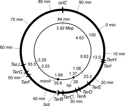
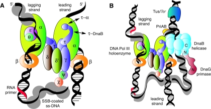
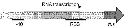
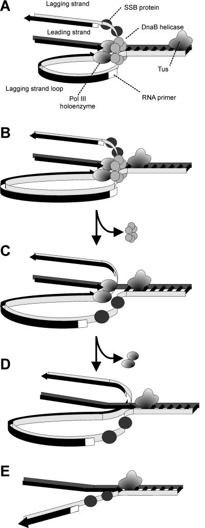
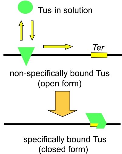
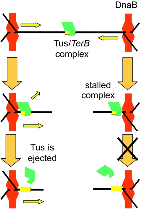
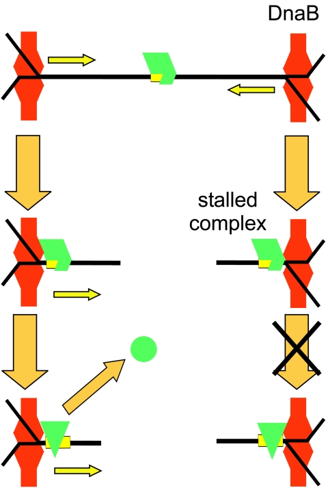
The arrest of DNA replication in Escherichia coli is triggered by the encounter of a replisome with a Tus protein-Ter DNA complex. A replication fork can pass through a Tus-Ter complex when traveling in one direction but not the other, and the chromosomal Ter sites are oriented so replication forks can enter, but not exit, the terminus region. The Tus-Ter complex acts by blocking the action of the replicative DnaB helicase, but details of the mechanism are uncertain. One proposed mechanism involves a specific interaction between Tus-Ter and the helicase that prevents further DNA unwinding, while another is that the Tus-Ter complex itself is sufficient to block the helicase in a polar manner, without the need for specific protein-protein interactions. This review integrates three decades of experimental information on the action of the Tus-Ter complex with information available from the Tus-TerA crystal structure. We conclude that while it is possible to explain polar fork arrest by a mechanism involving only the Tus-Ter interaction, there are also strong indications of a role for specific Tus-DnaB interactions. The evidence suggests, therefore, that the termination system is more subtle and complex than may have been assumed. We describe some further experiments and insights that may assist in unraveling the details of this fascinating process.


Figures

FIG. 1.
FIG. 1.
Positions of Ter sites and the tus gene on the E. coli chromosome. All Ter sites are oriented so that the replication forks can travel in the origin-to-terminus direction but not the opposite direction. The tus gene is just downstream of TerB.
FIG. 2.
FIG. 2.
Protein-protein interactions in the Escherichia coli replisome as it approaches the Tus-Ter termination block. (A) The DNA polymerase III (Pol III) holoenzyme is an asymmetric dimer containing 10 different subunits that include the twin polymerase (α) subunits that simultaneously replicate the two strands of the DNA template. (B) The replisome is a multiprotein complex made up of the DnaB helicase, the DnaG primase, and the Pol III holoenzyme. Each replicated strand commences with a short RNA primer synthesized by DnaG primase recruited from solution by interaction with DnaB. Single-stranded DNA is protected by SSB. Adapted from Fig. 2 of reference with the permission of the authors.
FIG. 3.
FIG. 3.
Nucleotide sequences of Ter sites from the E. coli chromosome and R6K plasmids. Base pairs that interact with the Tus protein are indicated by the shaded regions. In the orientation shown for these sequences, replication forks approaching from the left are blocked, while those entering from the right are unimpeded.
FIG. 4.
FIG. 4.
Relationship between TerB and the tus gene. The tus gene and its −10 promoter region and ribosome-binding site (RBS) are shown. The Tus protein regulates tus gene expression by binding to the TerB sequence and blocking the initiation of transcription of tus. The TerB sequence is enclosed in the box, and base pairs that interact with Tus are shaded as in Fig. 3.
FIG. 5.
FIG. 5.
Replisome of E. coli and mechanism of replication fork arrest by a Tus-Ter complex. (A) The replisome moving along the DNA template approaches Tus, and the DnaB helicase assists primase to lay down the last lagging-strand primer. (B) DnaB helicase action isblocked by Tus, and DnaB dissociates from the template. (C) DNA polymerase III (Pol III) holoenzyme completes leading-strand synthesis up to the Tus-Ter complex and (D) synthesizes the last Okazaki fragment on the lagging strand, which will eventually be ligated by DNA ligase to the penultimate fragment following removal of its RNA primer by DNA polymerase I (not shown). (E) The holoenzyme then dissociates, leaving a Y-forked structure that is single stranded on the lagging strand near the Tus-Ter complex.
FIG. 6.
FIG. 6.
The crystal structure of the Tus-TerA complex, PDB code 1ECR (85). Three views of the Tus-Ter complex are shown. The top view is looking down the DNA from the nonpermissive face of the complex. The middle view is rotated 90° from the first to show the front of the complex. The bottom view, rotated a further 90°, is along the DNA from the permissive end of the complex. The permissive and nonpermissive faces are indicated in the middle view. The balls indicate the (5′) strands that would pass through the central channel of the DnaB helicase. Images of protein structures in this and succeeding figures were generated in SWISS-PDB VIEWER version 3.7 (http://ca.expasy.org/spdbv/) (56) and rendered using POV-RAY version 3.1g.watcom.win32 (www.povray.org).
FIG. 7.
FIG. 7.
Summary of contacts between Tus and TerA. Adapted from reference with permission of the publisher. Arrows show interactions between amino acid side chains and groups in the base pairs. Residues in the TerA oligonucleotide used for determination of the crystal structure were A4 to T18 on one strand and T19 to T5 on the other and are shown with boldface outlines. Dashed lines indicate possible interactions at the permissive end that were not seen in the crystal structure (see the text for details).
FIG. 8.
FIG. 8.
Sequence and secondary structure of the Tus protein (data are from reference 85). The 31 residues that make nonspecific contacts to the DNA backbone are in blue. The 17 residues that make direct or water-mediated specific contacts with the DNA bases are in red.
FIG. 9.
FIG. 9.
Summary of the results of footprinting studies by Sista et al. (158) and Gottlieb et al. (54). Arrows indicate protection from hydroxyl radical cleavage. Filled circles indicate protection from methylation by dimethyl sulfate. Open circles show enhanced methylation. The base pairs that interact with Tus are shaded as in Fig. 3.
FIG. 10.
FIG. 10.
The nonpermissive face of the Tus-Ter complex. Four equivalent views of this face are shown, highlighting the features that might come into contact with the DnaB helicase. (A) Secondary structure elements that could contact DnaB; (B) residues at the nonpermissive face of the complex, including Glu49; (C) space-filling representation colored by residue type (red, acidic; blue, basic; yellow, polar; gray, aliphatic); (D) charge distribution on the Tus surface at the nonpermissive face. Charge was calculated without the TerA DNA in place, using atomic charges and Poisson-Boltzman calculation as implemented in SWISS-PDB VIEWER version 3.7 (56).
FIG. 11.
FIG. 11.
Tus-DNA and Tus-Ter binding. The solution form of Tus binds nonspecifically to DNA and scans along the double helix searching for a Ter site. On finding a Ter site, a series of conformational changes leads to formation of the closed Tus-Ter complex.
FIG. 12.
FIG. 12.
Sequence alignment of some Tus and Tus-like proteins. An alignment of the Tus protein sequences from E. coli, Salmonella enterica serovar Typhimurium, Klebsiella pneumoniae subsp. ozaenae, and Yersinia pestis, along with sequences of Tus-like proteins from the R394 plasmid of S. enterica serovar Typhimurium and the Rts-1 plasmid of Proteus vulgaris, was carried out and colored using the default parameters in CLUSTAL_X (64). Essentially, residues are colored where more than a given percentage of residues belong to one class: cyan, aliphatic and hydrophobic residues; orange, basic residues; purple, acidic residues; green, neutral hydrogen bonding residues. All glycines are colored brown, and all prolines are colored yellow. Secondary structure elements from the Tus-Ter crystal structure are shown above the alignment. Residues that make DNA backbone contacts in the crystal structure are shown with a blue block above the alignment. Those residues that make sequence-specific contacts with the Ter DNA are shown with a red block. Tus and Tus-like proteins were identified using PSI-BLAST (4).
FIG. 13.
FIG. 13.
Reconstruction of model atomic resolution structures of the DnaB helicase with threefold (A and C) and sixfold (B and D) symmetries. The helicase would approach the Tus-Ter complex with the upper face in C and D. Atomic resolution structures of the T7 gene 4 helicase domain (green) (157) and the N-terminal domain of DnaB (blue) (46) were docked into electron density maps determined by electron microscopy. The arrows in A indicate regions of the helicase structure that penetrate the electron microscopy surface envelope in the compressed helicase domains, suggesting that additional conformational changes in the atomic structure are necessary to fit the electron microscopy map. In both the threefold and sixfold models, there is additional unfilled density between the helicase and N-terminal domain (D, red arrow) that is likely due to 51 residues of the linker region not accounted for in the atomic structures. The figure is reproduced from reference with permission from the Journal of Molecular Biology.
FIG. 14.
FIG. 14.
A “complete dissociation” model of Tus action. As shown, the permissive face of the Tus-Ter complex is on the left and the nonpermissive face is on the right. DnaB approaching the permissive face for replication comes into contact with the Tus-Ter complex, leading to complete dissociation of Tus. DnaB approaching the nonpermissive (fork-blocking) face is blocked from proceeding farther by the Tus-Ter complex.
FIG. 15.
FIG. 15.
A simple two-step model of Tus-Ter and DnaB interactions. (Left) DnaB approaching the permissive face of the Tus-Ter complex promotes the formation of the open, nonspecifically bound form of Tus, which may dissociate directly or slide along the DNA. If DnaB moves into the Ter site before Tus can return to the specifically bound closed form, then helicase activity continues. (Right) DnaB approaching from the nonpermissive face cannot promote the formation of the open form of the complex, and further DNA unwinding is blocked.

References

    1. Abhyankar, M. M., J. M. Reddy, R. Sharma, E. Büllesbach, and D. Bastia. 2004. Biochemical investigations of the control of replication initiation of plasmid R6K. J. Biol. Chem. 279:6711-6719. - PubMed
    1. Abhyankar, M. M., S. Zzaman, and D. Bastia. 2003. Reconstitution of R6K DNA replication in vitro using 22 purified proteins. J. Biol. Chem. 278:45476-45484. - PubMed
    1. Altschul, S. F., W. Gish, W. Miller, E. W. Meyers, and D. J. Lipman. 1990. Basic local alignment search tool. J. Mol. Biol. 215:403-410. - PubMed
    1. Altschul, S. F., T. L. Madden, A. A. Schäffer, J. Zhang, Z. Zhang, W. Miller, and D. J. Lipman. 1997. Gapped BLAST and PSI-BLAST: a new generation of protein database search programs. Nucleic Acids Res. 25:3389-3402. - PMC - PubMed
    1. Amin, A. A., and J. Hurwitz. 1992. Polar arrest of the simian virus 40 tumor antigen-mediated replication fork movement in vitro by the tus protein-terB complex of Escherichia coli. J. Biol. Chem. 267:18612-18622. - PubMed

Publication types

MeSH terms

LinkOut - more resources