Comparative Study
. 2005 Oct 5;24(19):3400-10.
doi: 10.1038/sj.emboj.7600809. Epub 2005 Sep 8.

Wound-healing defect of CD18(-/-) mice due to a decrease in TGF-beta1 and myofibroblast differentiation

Affiliations
Comparative Study

Wound-healing defect of CD18(-/-) mice due to a decrease in TGF-beta1 and myofibroblast differentiation

Thorsten Peters et al. EMBO J. .

Abstract

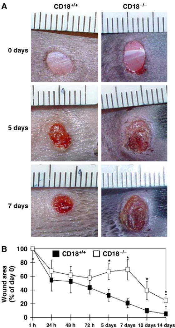
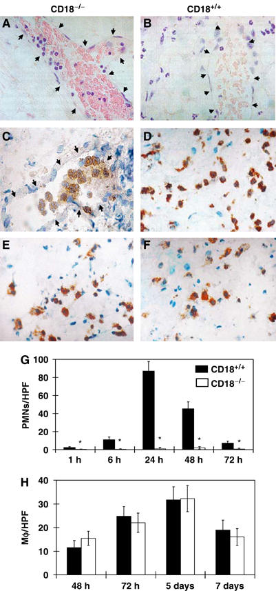
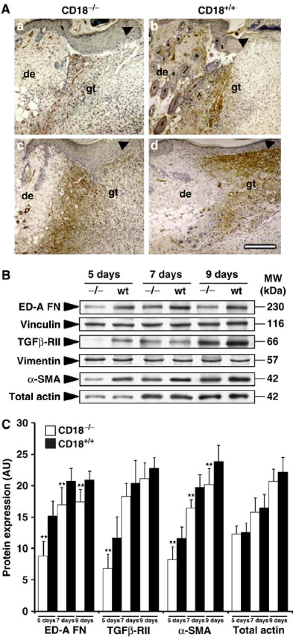
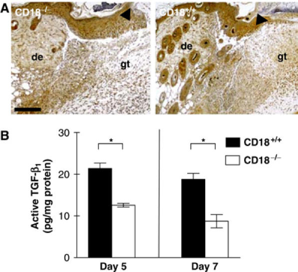
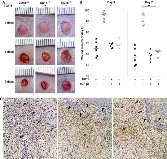
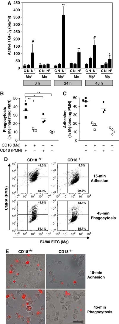
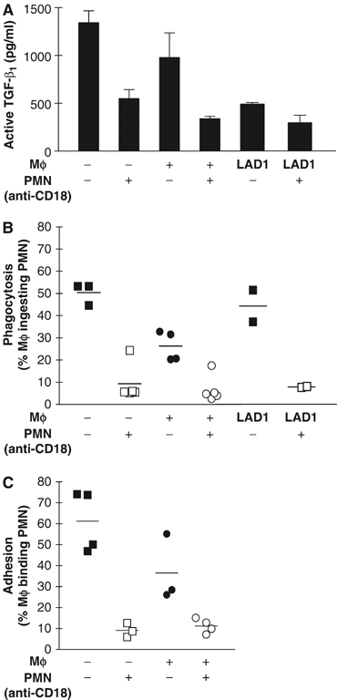
We studied the mechanisms underlying the severely impaired wound healing associated with human leukocyte-adhesion deficiency syndrome-1 (LAD1) using a murine disease model. In CD18(-/-) mice, healing of full-thickness wounds was severely delayed during granulation-tissue contraction, a phase where myofibroblasts play a major role. Interestingly, expression levels of myofibroblast markers alpha-smooth muscle actin and ED-A fibronectin were substantially reduced in wounds of CD18(-/-) mice, suggesting an impaired myofibroblast differentiation. TGF-beta signalling was clearly involved since TGF-beta1 and TGF-beta receptor type-II protein levels were decreased, while TGF-beta(1) injections into wound margins fully re-established wound closure. Since, in CD18(-/-) mice, defective migration leads to a severe reduction of neutrophils in wounds, infiltrating macrophages might not phagocytose apoptotic CD18(-/-) neutrophils. Macrophages would thus be lacking their main stimulus to secrete TGF-beta1. Indeed, in neutrophil-macrophage cocultures, lack of CD18 on either cell type leads to dramatically reduced TGF-beta1 release by macrophages due to defective adhesion to, and subsequent impaired phagocytic clearance of, neutrophils. Our data demonstrates that the paracrine secretion of growth factors is essential for cellular differentiation in wound healing.


Figures

Figure 1
Figure 1
Wound closure of full-thickness wounds is delayed in CD18−/− mice. Full-thickness (including the panniculus carnosus) excisional wounds were punched at two sites in the middle of the dorsum using 5-mm biopsy round knives. Each wound region was digitally photographed at the indicated time points, and wound areas were calculated using Adobe Photoshop® software. (A) Macroscopic observation of wounds in CD18−/− and WT mice. Representative results of six wounds in each cohort are shown. (B) Wound sizes at any given time point after wounding were expressed as percentage of initial (day 0) wound area for CD18−/− and WT mice. Results are expressed as the mean±s.d. (n=6). *P<0.05.
Figure 2
Figure 2
Impaired migration of PMN, but not Mφ, to wound beds of CD18−/− mice. H&E stainings (A, B) and immunostainings with a PMN-specific mAb (GR1) (C, D), as well as with an Mφ-specific mAb (F4/80) (E, F), were prepared from paraffin-embedded sections of CD18−/− (A, C, E) and WT mice (B, D, F) at 24 h (A–D) and 5 days (E, F) after full-thickness wounding. Arrows point at endothelial cells lining the vascular lumina. Subsequently, numbers of extravasated (G) GR1+ PMN and (H) F4/80+ Mφ within the dermis of the wound margins were counted in five high-power fields (HPF) at × 40 magnification using a light microscope. Results are given as the mean±s.d. (n=4). *P<0.05.
Figure 3
Figure 3
Reduced expression of myofibroblast-differentiation markers in wounds of CD18−/− mice. To detect key markers for myofibroblast differentiation and wound contraction, (A) paraffin-embedded granulation tissue of CD18−/− and WT mice was stained immunohistochemically for α-SMA, 5 (a, b) and 7 (c, d) days after wounding. The bar indicates 200 μm; de, adjacent dermis; gt, granulation tissue; arrows indicate epidermal leading edges. (B) Snap-frozen wound tissue was analysed by Western blotting, equilibrated to vimentin expression levels from Coomassie gels, to measure expression of myofibroblast markers including α-SMA, ED-A FN and TGFβ-RII at different time points. Panel B was assembled from separately performed blots for each indicated protein, using the same complete series of wound samples with identical loading between lanes (15 μg). (C) Semiquantitative balance analyses of Western blots were performed by densitometry of digitised Western blots. Data is given as the mean±s.d. **P<0.005.
Figure 4
Figure 4
Reduced TGF-β1 in the granulation tissue of CD18−/− mice. (A) Paraffin-embedded sections of wound sites obtained from CD18−/− and WT mice at day 5 after injury were stained immunohistochemically for TGF-β1 (brown). Whereas the typical staining pattern for TGF-β1 was observed in WT wounds that apart from an epidermal staining gave a strong signal in the wound bed-replenishing granulation tissue, CD18−/− wounds showed only weak staining in the granulation tissue located directly underneath the wound. The bar indicates 200 μm; de, adjacent dermis; gt, granulation tissue; arrows indicate epidermal leading edges. (B) Lysates of snap-frozen wound tissues from CD18−/− and WT mice obtained at the indicated time points after wounding were also prepared and subjected to ELISA to detect active TGF-β1. Data is given as the mean±s.d. *P<0.05.
Figure 5
Figure 5
Injection of TGF-β1 in wound margins rescues wound closure and myofibroblast differentiation in CD18−/− mice. Recombinant human TGF-β1 (‘TGF') was injected s.c. at four sites around the wound, allowing to infiltrate the wound margins, at a total dose of 0.45 μg per wound. Mock injections were made using only the solvent NaCl 0.9%. First injections were carried out on day 1 after wounding, followed by further injections every second day until wounds were harvested. (A) Macroscopic observation of wounds in CD18−/−, WT mice with or without injection of TGF-β1. (B) Wound sizes were assessed at the indicated time points after wounding as previously. Bars depict the median of each cohort. **P<0.005. (C) Paraffin-embedded granulation tissue of CD18−/− and WT mice was stained immunohistochemically for α-SMA 5 days after wounding. The bar indicates 100 μm; de, adjacent dermis; gt, granulation tissue; arrows indicate the newly formed epidermal leading edges.
Figure 6
Figure 6
Impaired in-vitro adhesion/phagocytosis of apoptotic PMN and concurrent deficiency of TGF-β1 release by Mφ under CD18-deficient conditions. To measure the uptake of apoptotic PMN by Mφ and TGF-β1 release that physiologically accompanies phagocytosis of apoptotic PMN by Mφ in the wound bed, CD18−/− or WT Mφ were cocultured with apoptotic CD18−/− (N) or WT (N+) PMN in an in vitro setting. As a control (C), Mφ and apoptotic PMN (not shown) were also incubated separately. (A) At the indicated time points, supernatants of cocultures were subjected to ELISA to detect active TGF-β1. Data is given as the mean±s.d. *,#P<0.05, **P<0.005. *,**Comparing CD18−/− and WT Mφ, #comparing CD18−/− and WT PMN. (B) In an identical setting, Mφ and PMN were coincubated, only for a shorter time. After 45 min, cells were collected from the well bottoms, stained and analysed by flow cytometry (for details, see Materials and methods in Supplementary data). Phagocytosis was assessed by calculating the percentage of Mφ phagocytosing PMN among the total Mφ counted (PMN-ingesting Mφ × 100/total number of Mφ). Bars indicate the median of each cohort. *P<0.05, **P<0.005. (C) Adhesion of apoptotic PMN preceding phagocytosis by Mφ depends on CD18. To investigate whether impaired phagocytosis in the absence of CD18 was due to an insufficient or absent adhesion between Mφ and PMN, we performed adhesion assays in which CD18−/− or WT Mφ were cocultured with apoptotic CD18−/− or WT PMN for 15 min. FACS-counted events were identified as single (i.e. nonadhering) apoptotic PMN (CMRA+ annexin-V+), single (nonadhering) Mφ (F4/80+) or as PMN–Mφ cell conjugates (CMRA+ F4/80+), stable enough to resist capillary shear forces during flow cytometry. The latter events were counted as Mφ-binding PMN (i.e. adhesion conjugates of Mφ with PMN), and were expressed as percentage of the total Mφ count (PMN-binding Mφ × 100/total number of Mφ). Each symbol indicates the median of a triplet analysis. *P<0.05, **P<0.005. (D) Representative flow-cytometric raw data of the adhesion and phagocytosis assays presented in (B, C) is provided. Mφ were stained using F4/80 FITC mAb; apoptotic PMN were loaded with CMRA prior to coculturing (for details see Materials and methods in Supplementary data). Cells appearing in the upper right quadrant of dot plots stain positive for both markers, thus representing either PMN adhering to Mφ after 15 min (‘Adhesion') or Mφ phagocytosing PMN after 45 min (‘Phagocytosis'), as also monitored by fluorescence microscopy. (E) Accordingly, Mφ phagocytosis of apoptotic PMN was assessed by immunofluorescence microscopy. For this purpose, coculturing of apoptotic PMN (labelled with CMRA, red/orange-fluorescing) with Mφ was performed using WT (left panel) and CD18−/− (right panel) PMN on Lab-Tek chamber slides (Nunc) for 15 min to detect adhesion, or for 45 min to assess phagocytosis. After cocultures, nonadherent PMN were washed away and microscopic pictures recorded digitally overlaying the differential interference contrast (DIC, using Nomarsky optics) with fluorescence pictures. The bars represent 40 μm.
Figure 7
Figure 7
Ablation of CD18 function leads to an impaired adhesion and phagocytosis of apoptotic human PMN with a reduced TGF-β1 release by Mφ. To assess uptake of apoptotic PMN and TGF-β1 release by Mφ in the human setting, monocytes were obtained from LAD1 patients and healthy human donors and differentiated in vitro to Mφ. PMN were isolated from the peripheral blood of healthy donors and rendered apoptotic by culturing for 20 h. To disrupt function of CD18 on PMN surface, PMN were preincubated with blocking mAbs against CD18 (clones IB4 or TS1/18) before they were added to Mφ, whereas control PMN with functional CD18 were left untreated. PMN were cocultured with Mφ (A) for 24 h to detect release of active TGF-β1, or (B) for 45 min to measure phagocytosis. (C) Adhesion of human PMN to Mφ was also assessed performing identical cocultures, but only for 15 min.

Similar articles

Cited by

References

    1. Anderson DC, Schmalstieg FC, Arnaout MA, Kohl S, Tosi MF, Dana N, Buffone GJ, Hughes BJ, Brinkley BR, Dickey WD, Abramson JS, Springer T, Boxer LA, Hollers JM, Smith CW (1984) Abnormalities of polymorphonuclear leukocyte function associated with a heritable deficiency of high molecular weight surface glycoproteins (GP138): common relationship to diminished cell adherence. J Clin Invest 74: 536–551 - PMC - PubMed
    1. Anderson DC, Smith CW (2001) Leukocyte Adhesion Deficiencies. New York, NY: McGraw-Hill
    1. Angele MK, Knoferl MW, Ayala A, Albina JE, Cioffi WG, Bland KI, Chaudry IH (1999) Trauma-hemorrhage delays wound healing potentially by increasing pro-inflammatory cytokines at the wound site. Surgery 126: 279–285 - PubMed
    1. Arnaout MA (1990) Structure and function of the leukocyte adhesion molecules CD11/CD18. Blood 75: 1037–1050 - PubMed
    1. Arora PD, McCulloch CA (1994) Dependence of collagen remodelling on alpha-smooth muscle actin expression by fibroblasts. J Cell Physiol 159: 161–175 - PubMed

Publication types

MeSH terms