Revascularization with saphenous vein bypasses for complex intracranial aneurysms
- PMID: 16148973
- PMCID: PMC1150875
- DOI: 10.1055/s-2005-870598
Revascularization with saphenous vein bypasses for complex intracranial aneurysms
Abstract
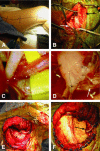
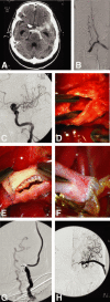
Most intracranial aneurysms can be managed with either microsurgical clipping or endovascular coiling. A subset of complex aneurysms with aberrant anatomy or fusiform/dolichoectatic morphology may require revascularization as part of a strategy that occludes the aneurysm or parent artery or both. Bypass techniques have been invented to revascularize nearly every intracranial artery. An aneurysm that will require a saphenous vein bypass is one that cannot be treated with conventional microsurgical clipping or endovascular coiling and also requires deliberate sacrifice of a major intracranial artery as part of the alternative treatment strategy. In the past 7 years the senior author (MTL) has performed a total of 110 bypasses, of which 46 were for aneurysms. Twenty-two of these patients received high-flow extracranial-to-intracranial bypasses using saphenous vein grafts, of which 16 had aneurysms that were giant in size. We review the indications for saphenous vein bypasses for complex intracranial aneurysms, surgical techniques, and clinical management strategies.
Figures


Similar articles
-
In situ bypass in the management of complex intracranial aneurysms: technique application in 13 patients.Neurosurgery. 2005 Jul;57(1 Suppl):140-5; discussion 140-5. doi: 10.1227/01.neu.0000163599.78896.f4. Neurosurgery. 2005. PMID: 15987580
-
Anterior cerebral artery bypass for complex aneurysms: an experience with intracranial-intracranial reconstruction and review of bypass options.J Neurosurg. 2014 Jun;120(6):1364-77. doi: 10.3171/2014.3.JNS132219. Epub 2014 Apr 18. J Neurosurg. 2014. PMID: 24745711 Review.
-
In situ bypass in the management of complex intracranial aneurysms: technique application in 13 patients.Neurosurgery. 2008 Jun;62(6 Suppl 3):1442-9. doi: 10.1227/01.neu.0000333808.64530.dd. Neurosurgery. 2008. PMID: 18695563
-
Combined microsurgical and endovascular management of complex intracranial aneurysms.Neurosurgery. 2003 Feb;52(2):263-74; discussion 274-5. doi: 10.1227/01.neu.0000043642.46308.d1. Neurosurgery. 2003. PMID: 12535354
-
Giant and complex aneurysms treatment with preservation of flow via bypass technique.Neurochirurgie. 2016 Feb;62(1):1-13. doi: 10.1016/j.neuchi.2015.03.008. Epub 2015 Jun 10. Neurochirurgie. 2016. PMID: 26072226 Review.
Cited by
-
Surgical repair of torn base of ruptured middle cerebral artery trifurcation aneurysm.Acta Neurochir (Wien). 2024 Mar 25;166(1):148. doi: 10.1007/s00701-024-06016-y. Acta Neurochir (Wien). 2024. PMID: 38523166 Free PMC article.
-
Stent infection and pseudoaneurysm formation after carotid artery stent treated by excision and in situ reconstruction with polytetrafluoroethylene graft: A case report.Surg Neurol Int. 2022 Jan 20;13:24. doi: 10.25259/SNI_1126_2021. eCollection 2022. Surg Neurol Int. 2022. PMID: 35127224 Free PMC article.
-
Magnetic resonance imaging flow quantification of non-occlusive excimer laser-assisted EC-IC high-flow bypass in the treatment of complex intracranial aneurysms.Clin Neuroradiol. 2012 Mar;22(1):39-45. doi: 10.1007/s00062-011-0116-z. Epub 2011 Dec 3. Clin Neuroradiol. 2012. PMID: 22138815
-
Extracranial-Intracranial Microsurgical Bypass Using a Y-Shaped Vein Graft From the Hand.Plast Surg (Oakv). 2024 May 6:22925503241249761. doi: 10.1177/22925503241249761. Online ahead of print. Plast Surg (Oakv). 2024. PMID: 39553519 Free PMC article.
-
Endovascular stenting of an extracranial-intracranial saphenous vein high-flow bypass graft: Technical case report.Surg Neurol Int. 2011;2:46. doi: 10.4103/2152-7806.79764. Epub 2011 Apr 19. Surg Neurol Int. 2011. PMID: 21660272 Free PMC article.
References
-
- Hopkins L N, Budny J L, Spetzler R F. Revascularization of the rostral brain stem. Neurosurgery. 1982;10:364–369. - PubMed
-
- Newell D W, Skirboll S L. Revascularization and bypass procedures for cerebral aneurysms. Neurosurg Clin N Am. 1998;9:697–711. - PubMed
-
- Spetzler R F, Carter L P. Revascularization and aneurysm surgery: current status. Neurosurgery. 1985;16:111–116. - PubMed
-
- Auguste K I, Quiñones-Hinojosa A, Lawton M T. The tandem bypass: subclavian artery-to-middle cerebral artery bypass with dacron and saphenous vein grafts. Technical case report. Surg Neurol. 2001;56:164–169. - PubMed
-
- Ausman J I, Diaz F G, Sadasivan B, Gonzeles-Portillo M, Jr, Malik G M, Deopujari C E. Giant intracranial aneurysm surgery: the role of microvascular reconstruction. Surg Neurol. 1990;34:8–15. - PubMed
LinkOut - more resources
Full Text Sources
Miscellaneous

