. 2005 May;15(2):133-41.
doi: 10.1055/s-2005-870599.

Superficial temporal artery to middle cerebral artery bypass

Affiliations

Superficial temporal artery to middle cerebral artery bypass

David W Newell. Skull Base. 2005 May.

Abstract

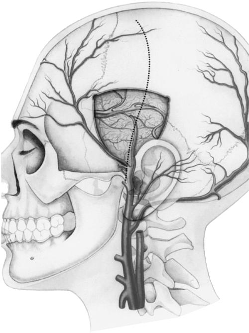
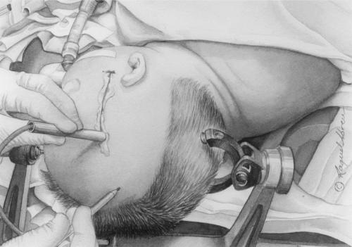
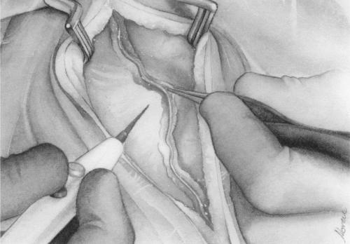
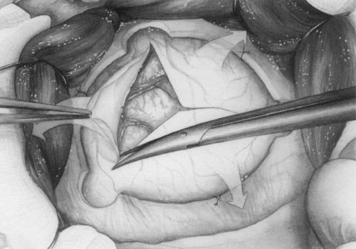
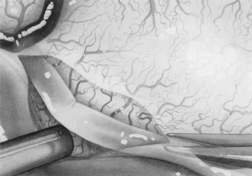
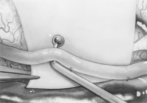
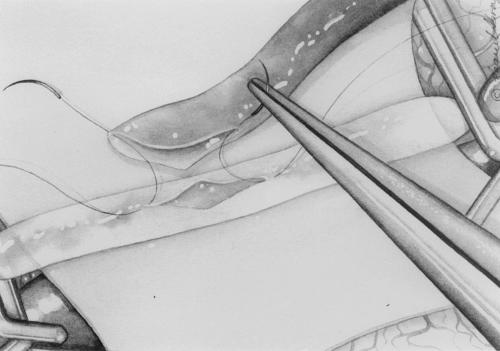
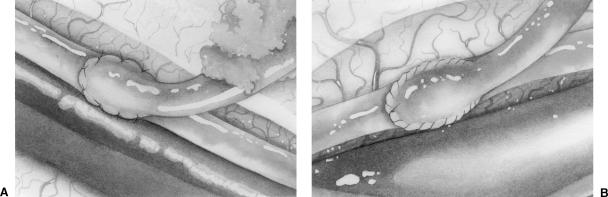
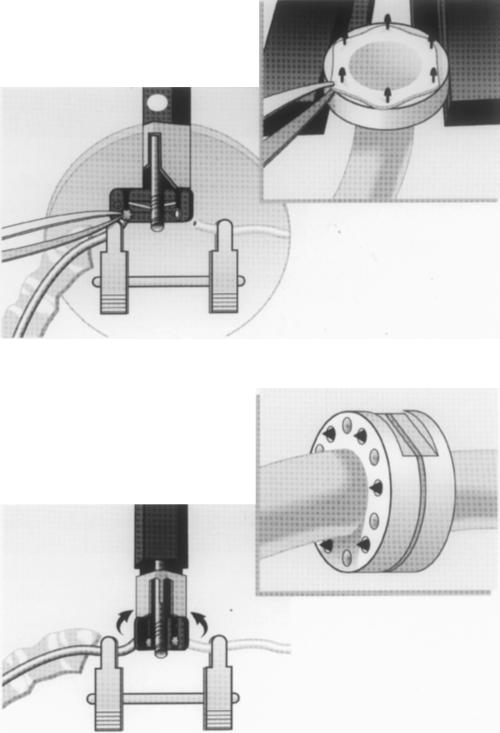
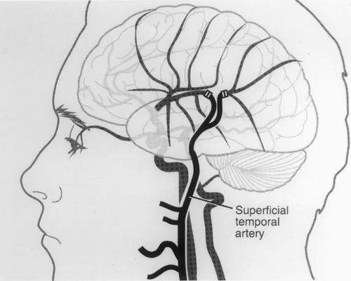
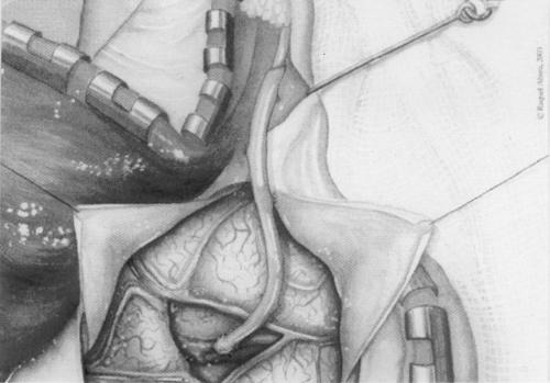
The superficial temporal artery to middle artery bypass is a technique that allows the blood supply from the extracranial carotid circulation to be routed to the distal middle cerebral artery branches. The procedure allows blood flow to bypass proximal lesions of the intracranial vasculature. The performance of this bypass requires specialized microvascular training and the use of microvascular techniques. The techniques involved in performing these procedures include microdissection of the superficial temporal artery in the scalp, microdissection of the recipient middle cerebral artery branches near the sylvian fissure, and anastomosis techniques using either microvascular sutures or a microanastomotic device. The successful completion of the bypass and subsequent patency requires meticulous attention to technical details.


Figures

Figure 1
Figure 1
Schematic representation of the STA-to-MCA anastomosis using the parietal branch as a donor artery. STA, superficial temporal artery; MCA, middle cerebral artery.
Figure 2
Figure 2
Photograph of an individual with a very prominent STA demonstrating the anatomy of the frontal and parietal branches (labeled). STA, superficial temporal artery.
Figure 3
Figure 3
The patient is positioned so the head can be turned to align the temporal and parietal region parallel to the floor. The parietal branch of the STA is identified using Doppler ultrasound and marked. STA, superficial temporal artery.
Figure 4
Figure 4
Illustration of the technique of dissection of the STA using needle-point cautery. Initial dissection of the scalp is performed directly over the artery and then the galeal layer is cauterized parallel to the artery to dissect it from the scalp and seal any small side branches. STA, superficial temporal artery.
Figure 5
Figure 5
Technique and location of dural opening illustrating the exposure of the cortex over the distal portion of the sylvian fissure. The graft is passed through the most inferior aspect of the dural opening.
Figure 6
Figure 6
Illustration of the technique of exposure of the cortical recipient branch of the MCA. The arachnoid layer is carefully teased off with a combination of sharp dissection and dissection with jeweler’s forceps. MCA, middle cerebral artery.
Figure 7
Figure 7
It is necessary to cauterize and divide multiple tiny perforating branches to the cortex to mobilize the recipient artery and place a small rubber working surface to perform the arteriotomy and anastomosis.
Figure 8
Figure 8
Placement of the end sutures aligns the donor and recipient arteries for the remainder of the anastomosis. 10–0 sutures with a noncutting needle are used.
Figure 9
Figure 9
This figure illustrates the two different options of suture closure of the anastomosis using either (A) interrupted or (B) running suture closure.
Figure 10
Figure 10
Illustration of the technique of end-to-end vessel anastomosis using the microanastomotic system. Each end of the vessel to be connected is loaded on the polyethelene ring and impaled on the six pins on each ring. The rings are then forced together and the pins of each ring insert into holes in the opposite ring and form a friction fit.
Figure 11
Figure 11
Schematic of the construction of a double limbed STA-MCA bypass from two branches of the STA to a divided branch of the distal MCA. STA, superficial temporal artery; MCA, middle cerebral artery.
Figure 12
Figure 12
Figure illustrating the course of a saphenous vein graft from the proximal STA (inferior) to a sylvian branch of the MCA (superior). STA, superficial temporal artery; MCA, middle cerebral artery.

Similar articles

Cited by

References

    1. Yasargil M G. A legacy of microneurosurgery: memoirs, lessons, and axioms. Neurosurgery. 1999;45:1025–1092. - PubMed
    1. Weerd A W de, Veldhuizen R J, Veering M M, Poortvliet D C, Jonkman E J. Long-term clinical and neurophysiological effects of reconstructive vascular surgery for cerebral ischemia. Acta Neurol Scand. 1989;79:311–315. - PubMed
    1. Diaz F G, Umansky F, Mehta B, et al. Cerebral revascularization to a main limb of the middle cerebral artery in the Sylvian fissure. An alternative approach to conventional anastomosis. J Neurosurg. 1985;63:21–29. - PubMed
    1. Gratzl O, Schmiedek P. STA-MCA bypass: results 10 years postoperatively. Neurol Res. 1983;5:11–18. - PubMed
    1. Spetzler R F. Retinal circulation after STA-MCA bypass. Neurosurgery. 1985;16:583. - PubMed