. 2005 Feb;15(1):71-82.
doi: 10.1055/s-2005-868164.

Cerebral revascularization in skull base tumors

Affiliations

Cerebral revascularization in skull base tumors

Stacey Quintero Wolfe et al. Skull Base. 2005 Feb.

Abstract

Skull base tumors involving the carotid artery pose a difficult surgical challenge. The potential for bypass grafting for cerebral revascularization carries inherent risks but may aid in tumor resection and control in those who warrant carotid sacrifice but have inappropriate natural cerebrovascular reserve. We include a review of the literature discussing the indications for carotid resection as part of skull base tumor surgery, indications for cerebral revascularization, balloon test occlusion, graft types and operative technique, complications, and results.


Figures

Figure 1
Figure 1
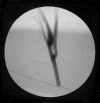
Balloon test occlusion: Right carotid angiogram with cross compression of left carotid. Apparent adequacy of collaterals through anterior communicating artery.
Figure 2
Figure 2
Single photon emission computed tomography-balloon test occlusion: Same patient as in Figure 1. Definite relative hypoperfusion of left hemisphere during balloon test occlusion.
Figure 3
Figure 3
Proximal saphenous vein graft harvesting from left thigh.
Figure 4
Figure 4
Pressure distention of saphenous vein graft. Testing for leaks and eliminating constrictive bands.
Figure 5
Figure 5
Distal saphenous vein graft—M2 end-to-side anastomosis completed.
Figure 6
Figure 6
Distended saphenous vein graft being prepared for tunneling to the neck.
Figure 7
Figure 7
Preauricular tunneling with chest tube.
Figure 8
Figure 8
Proximal saphenous vein graft-common carotid artery end-to-side anastomosis completed.
Figure 9
Figure 9
After completion of both anastomoses, flow reestablished through saphenous vein graft.
Figure 10
Figure 10
Cranial view of intraoperative angiogram prior to carotid sacrifice. Bypass is clearly patent.
Figure 11
Figure 11
Cervical view of intraoperative angiogram prior to carotid sacrifice. The saphenous vein graft is clearly seen anastomosed to the distal common carotid.
Figure 12
Figure 12
Bone flap closure. Sufficient craniectomy is created to avoid compression of the graft.
Figure 13
Figure 13
Final Doppler check of saphenous vein graft flow prior to leaving the operating room.

References

    1. Conley J. Free autogenous vein graft to the internal and common carotid arteries in the treatment of tumors of the neck. Ann Surg. 1953;137:205–214. - PMC - PubMed
    1. Lougheed W, Marshall B, Hunter M, et al. Common carotid to internal carotid bypass venous graft. Technical note. J Neurosurg. 1971;34:114–118. - PubMed
    1. Fisch U P, Olding D J, Senning A. Surgical therapy of internal carotid artery lesions of the skull base and temporal bone. Otolaryngol Head Neck Surg. 1980;88:548–554. - PubMed
    1. Glassock M, Smith P, Bond A, Whitaker S, Bartels L. Management of aneurysms of the petrous portion of the internal carotid artery by resection and primary anastomosis. Laryngoscope. 1983;93:1445–1453. - PubMed
    1. Sekhar L, Sen C, Jho H. Saphenous vein graft bypass of the cavernous internal carotid artery. J Neurosurg. 1990;72:35–41. - PubMed