. 2005 Sep;16(9):1097-109.
doi: 10.1089/hum.2005.16.1097.

Specific delivery of therapeutic RNAs to cancer cells via the dimerization mechanism of phi29 motor pRNA

Affiliations

Specific delivery of therapeutic RNAs to cancer cells via the dimerization mechanism of phi29 motor pRNA

Songchuan Guo et al. Hum Gene Ther. 2005 Sep.

Erratum in

  • Hum Gene Ther. 2006 Apr;17(4):476

Abstract

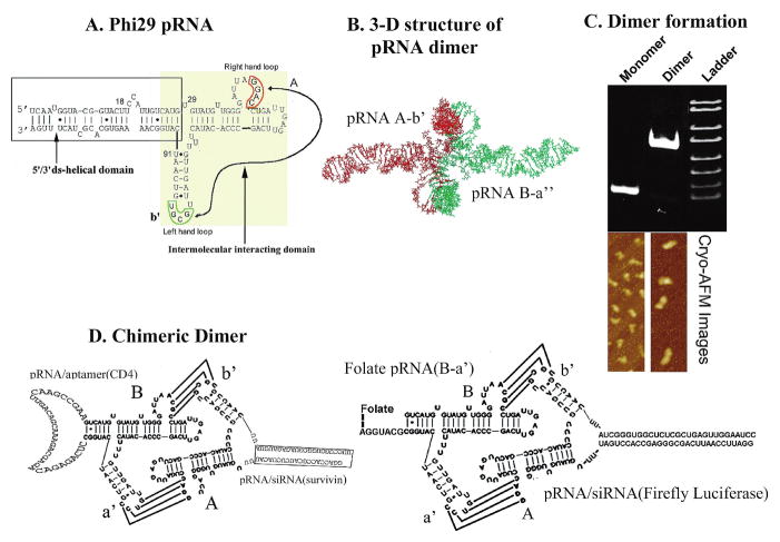
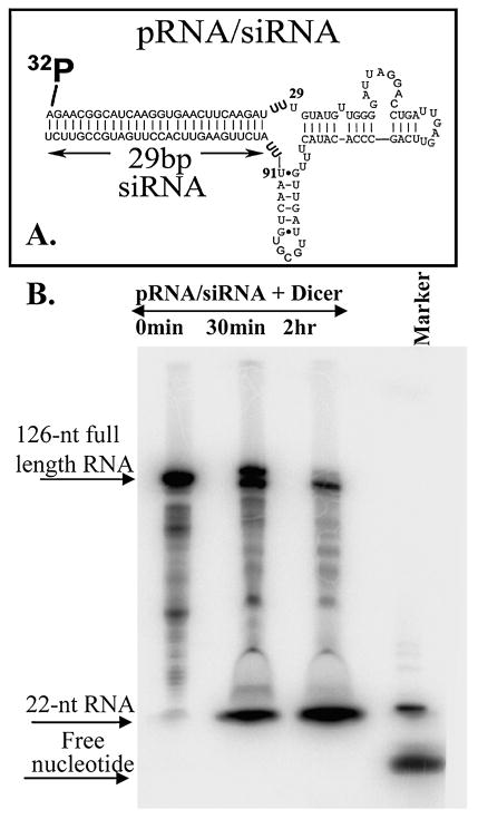
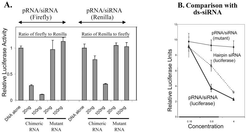
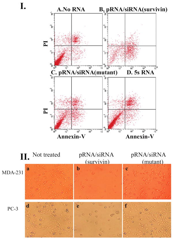
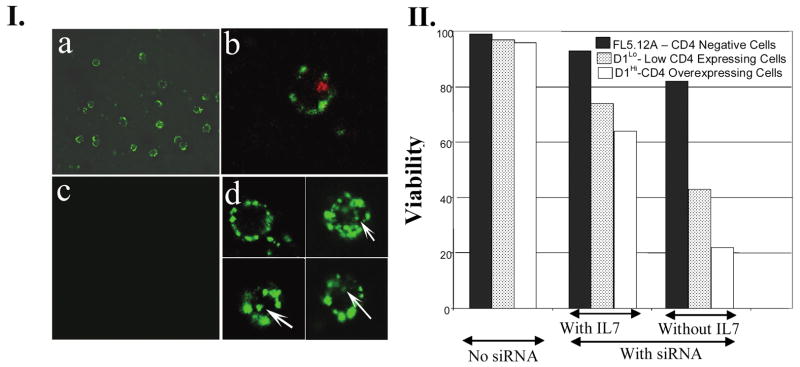
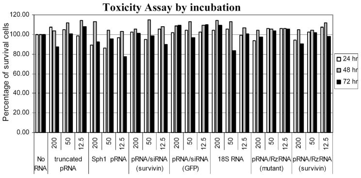
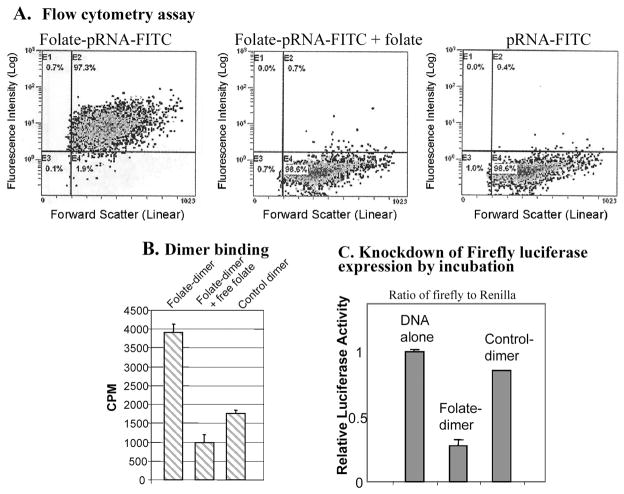
The application of small RNA in therapy has been hindered by the lack of an efficient and safe delivery system to target specific cells. Packaging RNA (pRNA), part of the DNA-packaging motor of bacteriophage phi29(Phi29), was manipulated by RNA nanotechnology to make chimeric RNAs that form dimers via interlocking right- and left-hand loops. Fusing pRNA with receptor-binding RNA aptamer, folate, small interfering RNA (siRNA), ribozyme, or another chemical group did not disturb dimer formation or interfere with the function of the inserted moieties. Incubation of cancer cells with the pRNA dimer, one subunit of which harbored the receptor-binding moiety and the other harboring the gene-silencing molecule, resulted in their binding and entry into the cells, and subsequent silencing of anti/proapoptotic genes. The chimeric pRNA complex was found to be processed into functional double-stranded siRNA by Dicer (RNA-specific endonuclease). Animal trials confirmed the suppression of tumorigenicity of cancer cells by ex vivo delivery. It has been reported [Shu, D., Moll, W.-D., Deng, Z., Mao, C., and Guo, P. (2004). Nano Lett. 4:1717-1724] that RNA can be used as a building block for bottom-up assembly in nanotechnology. The assembly of protein-free 25-nm RNA nanoparticles reported here will allow for repeated long-term administration and avoid the problems of short retention time of small molecules and the difficulties in the delivery of particles larger than 100 nm.


Figures

FIG. 1
FIG. 1
Sketch of sequence and structure of pRNA chimeras. (A) φ29 pRNA sequence and secondary structure. The right- and left-hand loops are circled in orange and green, respectively. The double-stranded helical domain on the 5′/3′ ends is framed, and the domain for dimer formation is shaded. The curved line points to the two interacting loops. (B) Three-dimensional structure of pRNA dimer. (C) Native polyacrylamide gel showing monomer and dimer of the pRNA chimeras exhibiting different migration rates. Below the gel are cryo-AFM images of 29 pRNA monomer and dimer. The colors reflect the thickness and height of the molecule; the brighter the color, the thicker or taller the molecule. (D) Design of chimeric pRNA dimers harboring foreign moieties (see Nomenclature of RNA Subunits, under Results).
FIG. 2
FIG. 2
Processing of chimeric pRNA/siRNA complex by Dicer. The structures of pRNA/siRNA and pRNA vector are shown in (A) and (B). Processing of pRNA/siRNA into 22-bp siRNAs by recombinant Dicer. The chimeric pRNA/siRNA with 5′-end 32P labeling was incubated with purified recombinant Dicer for 30 min and 2 hr, respectively, and then separated on a denaturing PAGE/urea gel. A radiolabeled 22-nucleotide RNA was used as a molecular weight marker.
FIG. 3
FIG. 3
Functional assay of chimeric pRNA/siRNA(GFP) by transfection. (AC) Fluorescence microscopy images showing the silencing of GFP gene by transfection. (A) Dose-dependent silencing of GFP gene by chimeric pRNA/siRNA(GFP) (left column). A mutant pRNA/siRNA (right column) served as negative control. (B) GFP expression of cells transfected with various RNAs: (a) no RNA; (b) synthesized double-stranded siRNA(GFP); (c) double-stranded siRNA(LacZ) control; (d) pRNA/siRNA(GFP); (e) pRNA/siRNA(mutant); (f) pRNA vector alone. (C) Comparison of the performance of (a) chimeric pRNA/siRNA(GFP) and (b) conventional double-stranded siRNA(GFP) at the same molar concentration; (c) control with no siRNA treatment. (D) Northern blot to examine the effect of chimeric pRNA/siRNA(GFP) on GFP mRNA level after transfection. Lanes 1 and 2 show the effects of two different constructs of pRNA/siRNA(GFP); lane 3, double-stranded siRNA; lane 4, cells without RNA treatment. rRNA was used as loading control.
FIG. 4
FIG. 4
Functional assay of chimeric pRNA/siRNA targeting luciferase by transfection. (A) Dual reporter luciferase assay showing the specific knockdown of firefly luciferase or Renilla luciferase expression by pRNA/siRNA(firefly) or pRNA/siRNA(Renilla), respectively, in a dose-dependent manner. (B) Comparison of the activities of conventional hairpin siRNA(luciferase) and pRNA/siRNA(luciferase). pRNA/siRNA(mutant) with mutations in siRNA sequences was included as a nonspecific control.
FIG. 5
FIG. 5
Apoptosis and cell death induced by transfection of chimeric pRNA harboring siRNA targeting survivin. (I) Breast cancer MCF-7 cells were transfected with pRNA/siRNA(survivin) and apoptosis was monitored by PI–annexin V double-labeling followed by flow cytometry. Cells in the bottom right quadrant represent apoptotic cells. (II) Breast cancer cells (MDA-231) and prostate cancer cells (PC-3) were transfected with 20 pmol of pRNA/siRNA(survivin) in 24-well plates and images were taken 24 hr after transfection. The mutant pRNA/siRNA was transfected in parallel as a negative control.
FIG. 6
FIG. 6
Functional assay of pRNA/siRNA chimera targeting proapoptotic factor BAD. (A) pRNA/siRNA(BAD) and control siRNAs (10 nM) were introduced into pro-B cells by electroporation, combined with a transfection reagent on day 1. Cells were washed to remove IL-3 on day 2 and assayed for viability on day 3. (B) BAD protein levels were compared in cells transfected with chimeric siRNA(BAD) or two mutant controls containing different mutations within siRNA sequences. Control cells were treated with pRNA alone. Cell lysates were prepared (Khaled et al., 2001) on day 3 and proteins were separated by 12% SDS–PAGE followed by Western blot with BAD antibody (Cell Signaling Technology, Beverly, MA). Numbers below the panel indicate the remaining BAD level, expressed as a percentage compared with control cells. (C) Pro-B cells transfected with pRNA/siRNA(survivin) or mutant were grown in complete medium containing IL-3 or deprived of IL-3. The impact of RNA on cell morphology was observed by microscopy.
FIG. 7
FIG. 7
Specific delivery of chimeric pRNA/siRNA by CD4 receptor. (I) Green circles corresponding to the binding of FITC-labeled pRNA dimer containing CD4-binding aptamer to lymphocytes were shown by confocal microscopy (a) and the entry of RNA was shown as a green spot inside the cell (d). Texas Red-labeled transferrin was used as a positive control of internalization (b). No binding was observed in the control cell line without CD4 expression (c). (II) Incubation of RNA dimer led to the specific suppression of cell viability, as measured by trypan blue exclusion assay. Three different cell lines with no, low, or high CD4 expression level were incubated with the pRNA/siRNA(survivin)-pRNA/aptamer(CD4) complex in the presence or absence of IL-7.
FIG. 8
FIG. 8
Toxicity assay of chimeric pRNA targeting survivin. HeLaT4 cells were seeded in 24-well plates so that they will be 30–50% confluent at the time of incubation. Twenty-four hours after seeding, the indicated amount of RNA was added into each well. Cells were further incubated in incubator for 24, 48, and 72 hr and the number of viable cells was counted by hemacytometer. The relative survival rates displayed were obtained by dividing the number of surviving cells in each treatment by the number of surviving cells without treatment with RNA.
FIG. 9
FIG. 9
Specific delivery of chimeric pRNA/siRNA by folate-pRNA. (A) Flow cytometry analyses of the binding of FITC-labeled folate-pRNA to KB cells. Left: Cells were incubated with folate-pRNA labeled with FITC. Middle: Cells were preincubated with free folate, which served as a blocking agent to compete with folate-pRNA for binding to the receptor. Right: Binding was also tested using folate-free pRNA labeled with FITC as a negative control. The percentages of FITC-positive cells are shown in the top right quadrants. (B) Specific binding of folate-pRNA dimer to KB cells. After incubation of cells with the [3H]folate-pRNA dimer in the presence (middle column) or absence (left column) of free folate, cells were isolated and subjected to scintillation counting. The right-hand column represent 3H-labeled dimer without folate labeling as a negative control. (C) In a knockdown assay by incubation, folate-chimeric dimer complex containing pRNA(B-a′)/folate and pRNA(A-b′)/siRNA(firefly) was incubated with KB cells for 3 hr to allow the binding and entry of RNA. The luciferase level was measured the next day in the dual reporter system. The control dimer was identical to the folate dimer except for its lack of folate labeling.
FIG. 10
FIG. 10
The potential use of pRNA hexamers as polyvalent gene delivery vectors. Six copies of pRNA have been found to form a hexameric ring to drive the DNA-packaging motor of bacterial virus φ29. There would therefore be six positions available to carry foreign moieties for targeting, therapy, and detection.

References

    1. AKKINA R, BANERJEA A, BAI J, ANDERSON J, LI MJ, ROSSI J. siRNAs, ribozymes and RNA decoys in modeling stem cell-based gene therapy for HIV/AIDS. Anticancer Res. 2003;23:1997–2005. - PubMed
    1. BLOUNT KF, UHLENBECK OC. The hammerhead ribozyme. Biochem Soc Trans. 2002;30:1119–1122. - PubMed
    1. BRUMMELKAMP TR, BERNARDS R, AGAMI R. A system for stable expression of short interfering RNAs in mammalian cells. Science. 2002;296:550–553. - PubMed
    1. CARMICHAEL GG. Medicine: Silencing viruses with RNA. Nature. 2002;418:379–380. - PubMed
    1. CHEN C, ZHANG C, GUO P. Sequence requirement for hand-in-hand interaction in formation of pRNA dimers and hexamers to gear φ29 DNA translocation motor. RNA. 1999;5:805–818. - PMC - PubMed

Publication types

MeSH terms