. 2005 Dec 15;392(Pt 3):467-74.
doi: 10.1042/BJ20051319.

Role for a P-type H+-ATPase in the acidification of the endocytic pathway of Trypanosoma cruzi

Affiliations

Role for a P-type H+-ATPase in the acidification of the endocytic pathway of Trypanosoma cruzi

Mauricio Vieira et al. Biochem J. .

Abstract

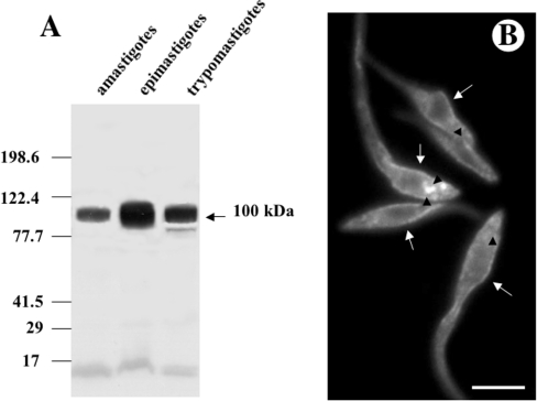
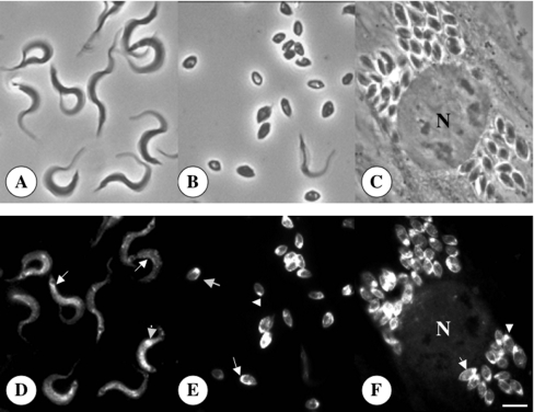
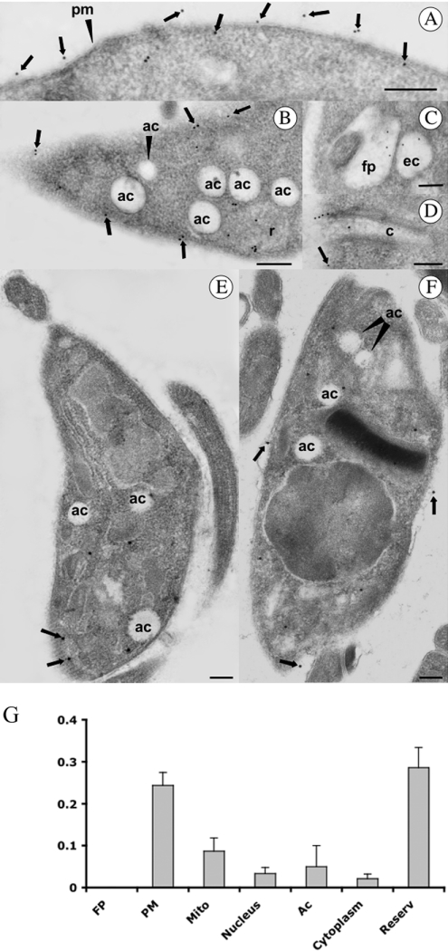
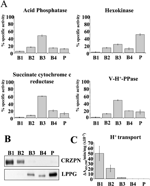
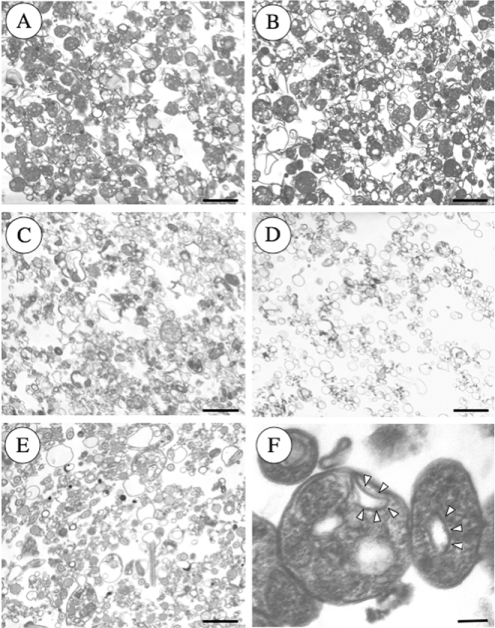
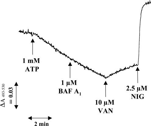
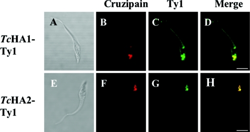
Previous studies in Trypanosoma cruzi, the etiologic agent of Chagas disease, have resulted in the cloning and sequencing of a pair of tandemly linked genes (TcHA1 and TcHA2) that encode P (phospho-intermediate form)-type H+-ATPases with homology to fungal and plant proton-pumping ATPases. In the present study, we demonstrate that these pumps are present in the plasma membrane and intracellular compartments of three different stages of T. cruzi. The main intracellular compartment containing these ATPases in epimastigotes was identified as the reservosome. This identification was achieved by immunofluorescence assays and immunoelectron microscopy showing their co-localization with cruzipain, and by subcellular fractionation and detection of their activity. ATP-dependent proton transport by isolated reservosomes was sensitive to vanadate and insensitive to bafilomycin A1, which is in agreement with the localization of P-type H+-ATPases in these organelles. Analysis by confocal immunofluorescence microscopy revealed that epitope-tagged TcHA1-Ty1 and TcHA2-Ty1 gene products are localized in the reservosomes, whereas the TcHA1-Ty1 gene product is additionally present in the plasma membrane. Immunogold electron microscopy showed the presence of the H+-ATPases in other compartments of the endocytic pathway such as the cytostome and endosomal vesicles, suggesting that in contrast with most cells investigated until now, the endocytic pathway of T. cruzi is acidified by a P-type H+-ATPase.


Figures

Figure 1
Figure 1. Western blot analysis of T. cruzi H+-ATPase (A) and immunofluorescence microscopy showing the localization of TcHAs in epimastigotes of T. cruzi (B)
(A) Homogenates containing 10 μg of protein from amastigotes, epimastigotes and trypomastigotes were subjected to SDS/PAGE on 10% polyacrylamide gels and transferred to nitrocellulose membranes. Lanes were probed with affinity purified anti-TcHAf antibody. Migration positions of prestained molecular mass standards are shown to the left of the gel. (B) Epimastigotes were treated with the affinity-purified antibody against TcHAs. Labelling of the plasma membrane (arrows) and large vacuoles (arrowheads) is evident. Scale bar, 5 μm.
Figure 2
Figure 2. Immunofluorescence microscopy showing the localization of TcHAs in trypomastigote (D) and amastigote (E, F) forms of T. cruzi
Upper panels show the same cells as in lower panels by bright field microscopy. N, host cell nucleus. Scale bar, 4 μm (AF). Labelling of the plasma membrane (arrows) and intracellular structures (arrowheads) is evident in both trypomastigotes and amastigotes.
Figure 3
Figure 3. Immunocytochemical localization of H+-ATPase in epimastigotes (A–C), trypomastigote (E) and amastigote (F) stages of T. cruzi
Particles (15 nm) were used to localize the H+-ATPase. Ac, acidocalcisome; C, cytostome; EV, endocytic vacuole; FP, flagellar pocket; PM, plasma membrane; R, reservosome. Arrowheads show the plasma membrane localization. Scale bars, 0.5 μm. (G) The density of gold particles was determined in the FP, mitochondria (Mito), nucleus, acidocalcisomes (ac), cytosol (cytoplasm) and reservosomes (reserv). Higher densities were found in the plasma membrane and reservosomes. Results are expressed as mean±S.E.M.
Figure 4
Figure 4. Localization of TcHAs in epimastigotes by confocal laser scanning microscopy (B–D, H–J) and immunoelectron microscopy (E–F)
(BD) Fluorescence images of epimastigote forms probed with antibody against TcHAf in red (B and D), and antibody against cruzipain in green (C and D). (D) Overlay of (B) and (C) showing co-localization in yellow. (A) Differential interference contrast image of the same cell. Scale bar, 5 μm (AD). (E, F) Epimastigote forms probed with antibody against TcHAf (20 nm particles) and antibody against cruzipain (10 nm particles) labelling the reservosomes (R). Labelling of the plasma membrane with antibodies against TcHAf was also evident (arrow). Scale bars, 125 nm. (HJ) Fluorescence images of epimastigote forms probed with antibody against TcHAf (at a 1:50 dilution to detect only the reservosomes) in green (H and J) and antibody against the V-H+-ATPase in red (I and J). (J) Overlay of (H) and (I) showing lack of co-localization. Scale bar, 5 μm.
Figure 5
Figure 5. Distribution of H+-ATPase activity from epimastigotes on sucrose density gradients
ATP-dependent bafilomycin A1 (0.5 μM)-insensitive proton transport activity (C) is concentrated in fractions B1 and B2. Its distribution was compared with that of established organelle markers: acid phosphatase (lysosomes); hexokinase (glycosomes); succinate cytochrome c reductase (mitochondria), and vacuolar proton pyrophosphatase (V-H+-PPase, acidocalcisomes). Scale bars show mean±S.E.M. as percentage of total recovered activity (or total activity for H+-transport (C) in absorbance/min per mg protein from 4 experiments. (B) Western blot analysis showing that cruzipain (CRZPN), which concentrates in reservosomes and lipopeptidephosphoglycan (LPPG, a plasma membrane marker) are in the different fractions of the gradient. Anti-cruzipain reactivity is present in fractions B1 and B2 whereas LPPG reactivity is absent in these fractions.
Figure 6
Figure 6. Representative electron micrographs of the subcellular fractions of epimastigotes
Note that reservosomes are enriched in B1 (A) and B2 (B) fractions. B3 (C) and B4 (D) contain other vacuoles that resemble mitochondrial fragments and microsomes, whereas the pellet (E) fraction also contains flagella and acidocalcisomes. (F) Shows reservosomes of fraction B1 at higher magnification showing the presence of internal membrane-bound vesicles (arrowheads). Scale bars, 0.5 μm (AE) and 0.1 μm (F).
Figure 7
Figure 7. ATP-induced acidification of reservosomes measured by Acridine Orange uptake
In the experiment shown, 0.13 mg of reservosome protein (fraction B1) was added per ml of assay medium. ATP (1 mM), bafilomycin A1 (BAF A1, 1 μM), sodium o-vanadate (VAN, 10 μM) and nigericin (NIG, 2.5 μM) were added where indicated.
Figure 8
Figure 8. Epimastigote forms expressing TcHA1-Ty1 and TcHA2-Ty1 fusion proteins
Epimastigotes were transfected with expression constructs pTEX-TcHA1-Ty1 (AD) and pTEX-TcHA2-Ty1 (EH) respectively. Stable transformants were labelled with monoclonal antibody BB2 anti-Ty1 in green (C and G) and polyclonal antibody against cruzipain in red (B and F). TcHA1-Ty1 localized to the reservosomes and the plasma membrane whereas TcHA2-Ty1 localized only to the reservosomes. (D) and (H) are overlays of (B) and (C), and (F) and (G) respectively, showing co-localization in yellow. (A) and (E) are differential interference contrast images of the same cells. Scale bars, 5 μm.

References

    1. Urbina J. A., Docampo R. Specific chemotherapy of Chagas disease: controversies and advances. Trends Parasitol. 2003;19:495–501. - PubMed
    1. Luo S., Scott D. A., Docampo R. Trypanosoma cruzi H+-ATPase 1 (TcHA1) and 2 (TcHA2) genes complement yeast mutants defective in H+ pumps and encode plasma membrane P-type H+-ATPases with different enzymatic properties. J. Biol. Chem. 2002;277:44497–44506. - PubMed
    1. Axelsen K. B., Palmgren M. G. Evolution of substrate specificities in the P-type ATPase superfamily. J. Mol. Evol. 1998;46:84–101. - PubMed
    1. Møller J. V., Juul B., le Maire M. Structural organization, ion transport, and energy transduction of P-type ATPases. Biochim. Biophys. Acta. 1996;1286:1–51. - PubMed
    1. Monk B. C., Perlin D. S. Fungal plasma membrane proton pumps as promising new antifungal targets. Crit. Rev. Microbiol. 1994;20:209–223. - PubMed

Publication types