

Bicoid cooperative DNA binding is critical for embryonic patterning in Drosophila
- PMID: 16150708
- PMCID: PMC1201621
- DOI: 10.1073/pnas.0506462102
Bicoid cooperative DNA binding is critical for embryonic patterning in Drosophila
Abstract
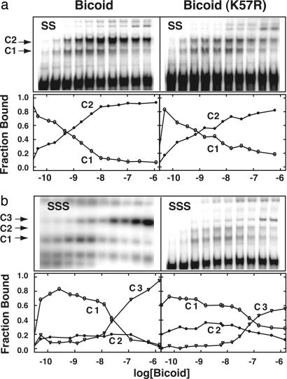
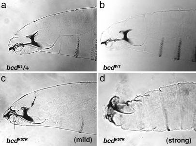
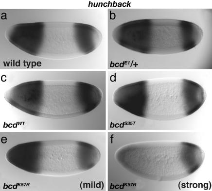
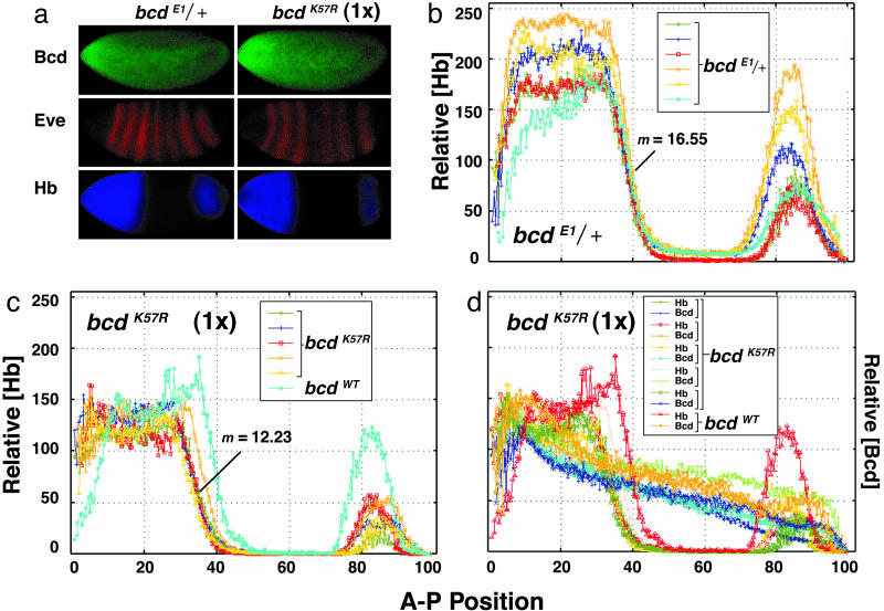
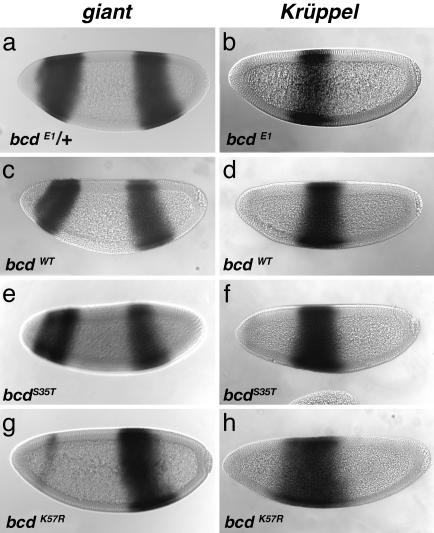
Cooperative interactions by DNA-binding proteins have been implicated in cell-fate decisions in a variety of organisms. To date, however, there are few examples in which the importance of such interactions has been explicitly tested in vivo. Here, we tested the importance of cooperative DNA binding by the Bicoid protein in establishing a pattern along the anterior-posterior axis of the early Drosophila embryo. We found that bicoid mutants specifically defective in cooperative DNA binding fail to direct proper development of the head and thorax, leading to embryonic lethality. The mutants did not faithfully stimulate transcription of downstream target genes such as hunchback (hb), giant, and Krüppel. Quantitative analysis of gene expression in vivo indicated that bcd cooperativity mutants were unable to accurately direct the extent to which hb is expressed along the anterior-posterior axis and displayed a reduced ability to generate sharp on/off transitions for hb gene expression. These failures in precise transcriptional control demonstrate the importance of cooperative DNA binding for embryonic patterning in vivo.
Figures
Similar articles
-
Spatial bistability generates hunchback expression sharpness in the Drosophila embryo.PLoS Comput Biol. 2008 Sep 26;4(9):e1000184. doi: 10.1371/journal.pcbi.1000184. PLoS Comput Biol. 2008. PMID: 18818726 Free PMC article.
-
Stable, precise, and reproducible patterning of bicoid and hunchback molecules in the early Drosophila embryo.PLoS Comput Biol. 2009 Aug;5(8):e1000486. doi: 10.1371/journal.pcbi.1000486. Epub 2009 Aug 28. PLoS Comput Biol. 2009. PMID: 19714200 Free PMC article.
-
3 minutes to precisely measure morphogen concentration.PLoS Genet. 2018 Oct 26;14(10):e1007676. doi: 10.1371/journal.pgen.1007676. eCollection 2018 Oct. PLoS Genet. 2018. PMID: 30365533 Free PMC article.
-
A matter of time: Formation and interpretation of the Bicoid morphogen gradient.Curr Top Dev Biol. 2020;137:79-117. doi: 10.1016/bs.ctdb.2019.11.016. Epub 2019 Dec 27. Curr Top Dev Biol. 2020. PMID: 32143754 Review.
-
Constraints and limitations on the transcriptional response downstream of the Bicoid morphogen gradient.Curr Top Dev Biol. 2020;137:119-142. doi: 10.1016/bs.ctdb.2019.12.002. Epub 2020 Jan 17. Curr Top Dev Biol. 2020. PMID: 32143741 Review.
Cited by
-
Spatial bistability generates hunchback expression sharpness in the Drosophila embryo.PLoS Comput Biol. 2008 Sep 26;4(9):e1000184. doi: 10.1371/journal.pcbi.1000184. PLoS Comput Biol. 2008. PMID: 18818726 Free PMC article.
-
A feed-forward relay integrates the regulatory activities of Bicoid and Orthodenticle via sequential binding to suboptimal sites.Genes Dev. 2018 May 1;32(9-10):723-736. doi: 10.1101/gad.311985.118. Epub 2018 May 15. Genes Dev. 2018. PMID: 29764918 Free PMC article.
-
Sequence-based model of gap gene regulatory network.BMC Genomics. 2014;15 Suppl 12(Suppl 12):S6. doi: 10.1186/1471-2164-15-S12-S6. Epub 2014 Dec 19. BMC Genomics. 2014. PMID: 25564104 Free PMC article.
-
Thermodynamics-based models of transcriptional regulation by enhancers: the roles of synergistic activation, cooperative binding and short-range repression.PLoS Comput Biol. 2010 Sep 16;6(9):e1000935. doi: 10.1371/journal.pcbi.1000935. PLoS Comput Biol. 2010. PMID: 20862354 Free PMC article.
-
The class E floral homeotic protein SEPALLATA3 is sufficient to loop DNA in 'floral quartet'-like complexes in vitro.Nucleic Acids Res. 2009 Jan;37(1):144-57. doi: 10.1093/nar/gkn900. Epub 2008 Nov 25. Nucleic Acids Res. 2009. PMID: 19033361 Free PMC article.
References
Publication types
MeSH terms
Substances
Grants and funding
LinkOut - more resources
Full Text Sources
Molecular Biology Databases
