. 2005 Dec;146(12):5176-87.
doi: 10.1210/en.2005-0811. Epub 2005 Sep 8.

Dehydroepiandrosterone restoration of growth hormone gene expression in aging female rats, in vivo and in vitro: evidence for actions via estrogen receptors

Affiliations

Dehydroepiandrosterone restoration of growth hormone gene expression in aging female rats, in vivo and in vitro: evidence for actions via estrogen receptors

Mary Iruthayanathan et al. Endocrinology. 2005 Dec.

Abstract

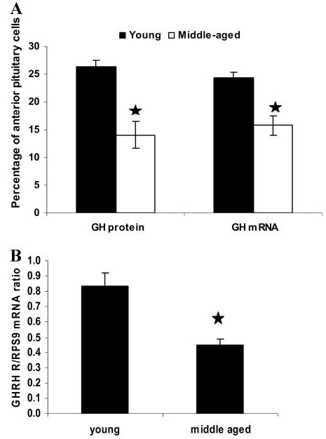
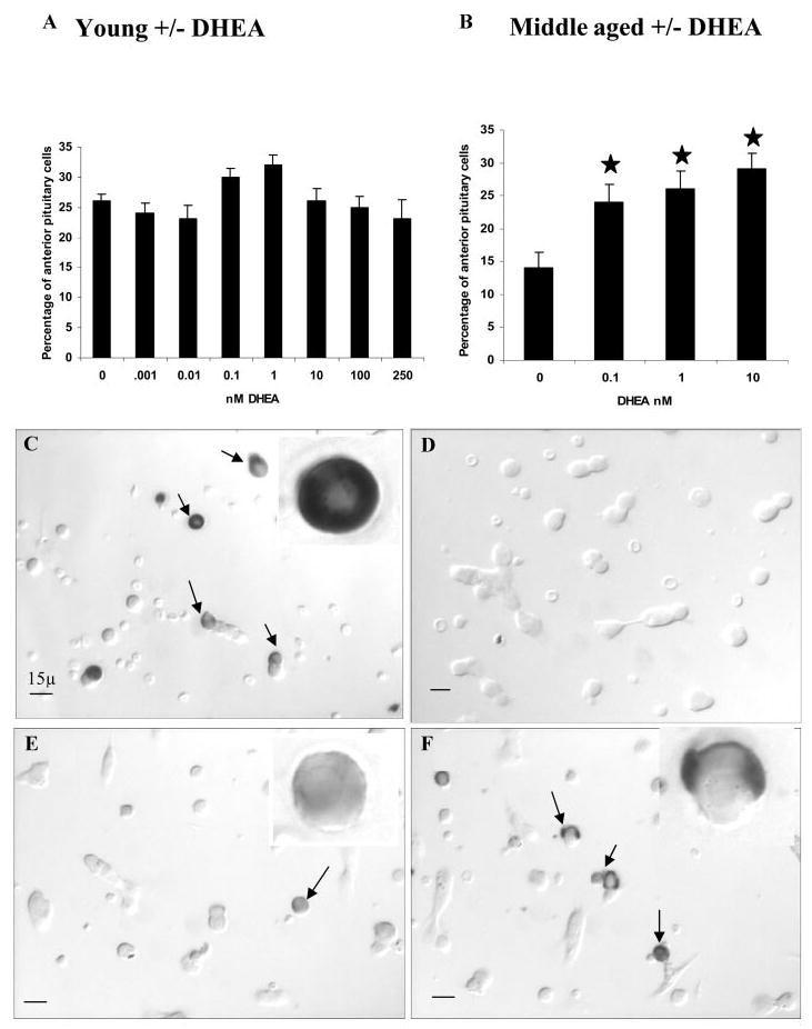
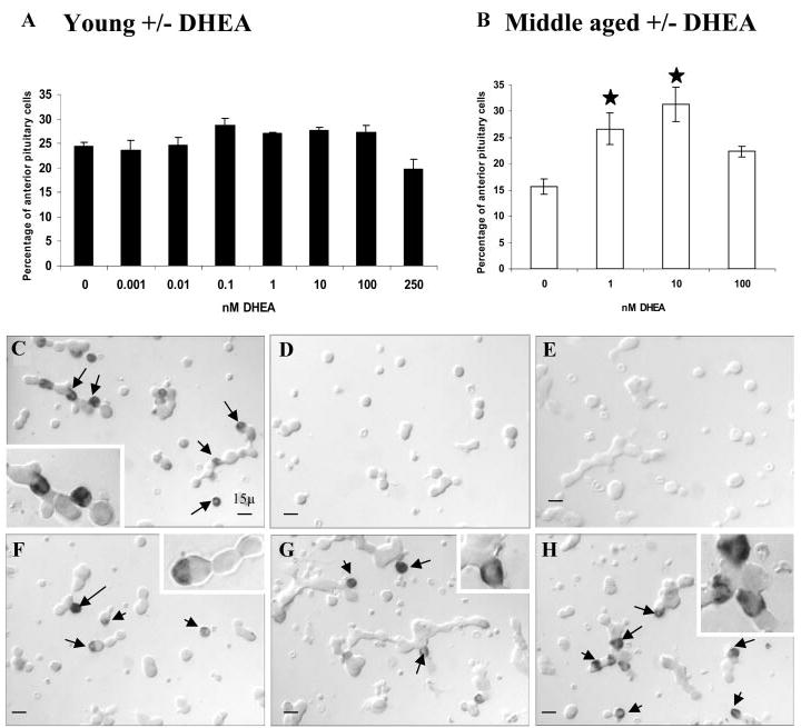
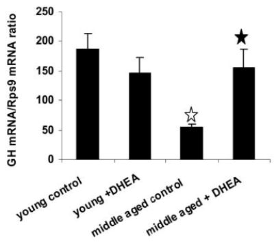
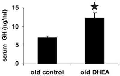
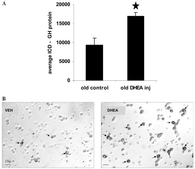
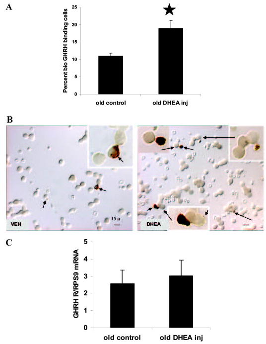
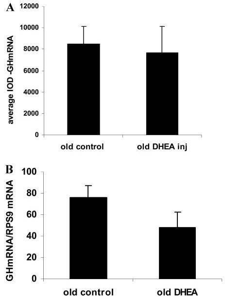
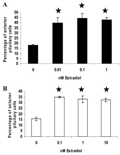
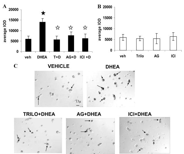
A decline in dehydroepiandrosterone (DHEA) and GH levels with aging may be associated with frailty and morbidity. Little is known about the direct effects of DHEA on somatotropes. We recently reported that 17beta-estradiol (E2), a DHEA metabolite, stimulates the expression of GH in vitro in young female rats. To test the hypothesis that DHEA restores function in aging somatotropes, dispersed anterior pituitary (AP) cells from middle-aged (12-14 months) or young (3-4 months) female rats were cultured in vitro with or without DHEA or E2 and fixed for immunolabeling or in situ hybridization. E2 increased the percentage of AP cells with GH protein or mRNA in the aged rats to young levels. DHEA increased the percentages of somatotropes (detected by GH protein or mRNA) from 14-16 +/- 2% to 29-31 +/- 3% (P < or = 0.05) and of GH mRNA (detected by quantitative RT-PCR) only in aging rats. To test DHEA's in vivo effects, 18-month-old female rats were injected with DHEA or vehicle for 2.5 d, followed by a bolus of GHRH 1 h before death. DHEA treatment increased serum GH 1.8-fold (7 +/- 0.5 to 12 +/- 1.3 ng/ml; P = 0.02, by RIA) along with a similar increase (P = 0.02) in GH immunolabel. GHRH target cells also increased from 11 +/- 1% to 19 +/- 2% (P = 0.03). Neither GH nor GHRH receptor mRNAs levels were changed. To test the mechanisms behind DHEA's actions, AP cells from aging rats were treated with DHEA with or without inhibitors of DHEA metabolism. Trilostane, aminogluthemide, or ICI 182,780 completely blocked the stimulatory effects of DHEA, suggesting that DHEA metabolites may stimulate aging somatotropes via estrogen receptors.


Figures

Fig. 1
Fig. 1
Decline in GH and GHRH R gene expression in the AP of middle-aged rats. A, Middle-aged female rats showed a significant decrease in the percentage (mean ± SEM) of somatotropes in AP detected by GH protein (immunocytochemistry) and GH mRNA (in situ hybridization) compared with young cycling diestrous rats.★, Statistical significance (P = 0.01; n = 3–6 rats). B, GHRH R mRNA was assayed by QRT-PCR and normalized by the housekeeping gene RPS9. Results are expressed as the ratio ± SEM. Middle-aged rats had significantly lower GHRH R mRNA (~50% of GHRH R mRNA found in young rats). ★, Significance (P = 0.02) compared with young control (young, n = 6; middle-aged, n = 4) rats.
Fig. 2
Fig. 2
Differential responses to DHEA by somatotropes (detected by GH protein) in young and middle-aged rats: Diestrous rat AP cells were grown in vitro for 24 h in vehicle or 0.001–250 nM DHEA (A). In parallel experiments, cells from middle-aged rats were grown in vehicle or 0.1, 1, and 10 nM DHEA (B). Cells were fixed and immunolabeled for GH. ★, Significance (P < 0.05) compared with aged vehicle controls (young, n = 6 animals; middle-aged, n = 6 animals). Illustrations of cells from the experiment described above showing immunolabeling from a young diestrous rat are shown in C. Immunolabeled cells from a middle-aged rat (E) and response to DHEA treatment with increased expression of GH protein-bearing cells are shown in F. Insets show a GH-immunolabeled cell from the field at a higher magnification. If anti-GH was absorbed with rat GH in the single-labeling protocol for GH, there was no label (D).
Fig. 3
Fig. 3
Differential responses to DHEA by somatotropes (detected by GH mRNA) in young and middle-aged rats. A, Cells from the AP of young diestrous rats treated with vehicle or DHEA (0.001–250 nM) for 24 h in vitro showed no change in the percentage of pituitary cells detected by GH mRNA (in situ hybridization). B, Cells from middle-aged animals, however, showed a significant increase (P < 0.05) in GH mRNA-expressing cells at 1 and 10 nM (young, n = 6; old, n = 3). Results are graphed as the mean ± SEM. Illustrations of fields from the experiment described above are shown in C–H. Arrows point to GH mRNA-labeled cells detected by in situ hybridization. C, Representative field from a young rat. A dose-dependent increase in GH mRNA-expressing AP cells in middle-aged rats with DHEA concentrations of 1 (G) and 10 (H) nM was seen compared with vehicle (F). Insets show some cells labeled for GH mRNA from the field at higher magnification. D and E, Control fields with no labeling in cells by the in situ hybridization protocol.
Fig. 4
Fig. 4
GH mRNA levels (determined by quantitative RT-PCR) in vehicle/DHEA-treated pituitary cells from young and middle-aged rats. GH mRNA was normalized with two housekeeping genes, RPS9 and HPRT (data not shown). Results are expressed as the ratio ± SEM. Middle-aged animals had significantly lower GH mRNA levels (~30% of GH mRNA in young rats), and DHEA (1 nM) treatment increased levels to those in young rats. ☆, Significance (P < 0.05) compared with young controls; ★, significance (P < 0.05) when DHEA treatment group was compared with corresponding vehicle treatment group (young, n = 5; middle-aged, n = 5 rats).
Fig. 5
Fig. 5
DHEA treatment in vivo enhances GH secretion. GH was assayed by RIA from trunk blood collected at the time of death. Average serum GH levels (nanograms per milliliter) ± SEM for vehicle-and DHEA-treated rats are shown in the graph. DHEA-injected 18-month-old female rats showed a significant increase (P = 0.02; ★) in serum GH levels in response to a bolus of GHRH injection compared with vehicle-injected control animals (vehicle-treated, n = 3 animals; DHEA-treated, n = 4).
Fig. 6
Fig. 6
DHEA treatment in vivo enhanced GH protein expression in the AP. A, Short-term in vivo administration of DHEA in aged female rats produced a significant increase in GH protein in AP (P = 0.02). GH protein was quantified by measuring IOD on digitized images of GH immunolabeling from dispersed pituitary cells from vehicle- or DHEA-treated aged rats (vehicle-treated, n = 3 animals; DHEA-treated, n = 4). Results are expressed as the average of the sum of the IODs ± SEM from 15 individual fields of individual animals.★, Statistical significance. B, Digitized images from the experiment described above illustrating the effects of DHEA on GH protein expression in the AP. Immunolabeling in a vehicle-injected control animal is shown (left panel). Arrows indicate GH cells identified by their protein. In vivo DHEA administration increased the expression of GH protein in the pituitary cells (right panel).
Fig. 7
Fig. 7
DHEA treatment increases GHRH-binding pituitary cells. A, Cells from AP of vehicle- or DHEA-treated rats were dispersed, plated for 1 h, and treated with biotinylated GHRH (1 nM) for 10 min before fixation. Affinity cytochemistry detecting the biotinylated ligand revealed a significant increase (P = 0.03; ★) in GHRH binding AP cells obtained from DHEA-treated aged rats. The graph represents the mean ± SEM (n = 3–4). B, Illustrations of double labeling (as described in Materials and Methods), showing GHRH-binding somatotropes in vehicle- and DHEA-treated aged female rats. Arrows show patchy black labeling of GHRH-binding sites on the cell membrane detected by affinity cytochemistry. Orange label indicates GH cells detected by GH protein (immunolabeling). DHEA-treated rats (right panel) had significantly more GHRH-bound pituitary cells than vehicle-treated control animals (left panel). The inset shows some of the GHRH-bound cells at higher magnification. Black label, GHRH binding; orange label, GH. C, Effect of DHEA on GHRH R mRNA levels in vivo. Half of the AP was collected for RNA extraction immediately after removal from the rat. GHRH R mRNA levels from the pituitary tissue quantified by real-time QRT-PCR, using RPS9 as internal standard, showed no changes in GHRH R mRNA levels between vehicle- and DHEA-injected aged rats. Results are expressed as the mean ± SEM (n = 3).
Fig. 8
Fig. 8
Effect of DHEA treatment in vivo on GH mRNA detected by in situ hybridization and quantitative RT-PCR. A, Cells from half of the AP of aged rats, injected with vehicle or DHEA (vehicle-treated, n = 3 animals; DHEA-treated, n = 4), were dispersed and plated on coverslips and fixed after 1 h to allow for adequate adhesion. The cells were labeled for GH mRNA by in situ hybridization, and the IOD was measured (as described in Materials and Methods) to report changes, if any, in GH mRNA levels. Results are graphed as the mean ± SEM and show no significant change in GH mRNA levels when detected by in situ hybridization. B, Half of the AP was collected for RNA extraction immediately after removal from the rat. The average GH mRNA levels ± SEM (n = 3) from the pituitary tissue quantified by real time QRT-PCR, using RPS9 as internal standard, showed no changes in GH mRNA levels between vehicle- and DHEA-injected aged rats.
Fig. 9
Fig. 9
Response of somatotropes (detected by GH protein and mRNA) to estrogen in middle-aged rats in vitro. Cells from middle-aged (12–14 months) rats were grown in vehicle or 0.01–10 nM E2. Cells were fixed and subjected to immunolabeling (A) or in situ hybridization (B) for GH. E2 in doses of 0.01–10 nM produced a rise in GH cells to the normal basal levels seen in young animals. The graph represents the mean ± SEM. ★, Significance (P < 0.05) in estrogen-treated middle-aged animals compared with their respective vehicle controls (n = 3–4 middle-aged rats).
Fig. 10
Fig. 10
Mechanism of DHEA action on aging somatotropes. A, Pituitary cells from middle-aged animals were grown for 24 h in vitro with 10 nM DHEA (D), with or without inhibitors [trilostane (T), aminogluthemide (AG), and ICI 182,780 (ICI)] in equimolar concentrations. The IOD of immunolabeled GH cells (see Materials and Methods) showed a significant increase in GH protein expression in the DHEA-treated group compared with vehicle treatment (P = 0.009;★). Trilostane (3β-HSD enzyme blocker) and aminoglutethemide (aromatase inhibitor) completely blocked the restorative effects of DHEA on aging somatotropes (P = 0.01 and P = 0.03, respectively). The ER antagonist ICI 182,780 also significantly blocked the DHEA effect (P = 0.01). The results are graphed as the mean ± SEM in A (n = 3). ☆, Significance compared with DHEA-treated group. B, The inhibitors themselves did not show any significant difference from the vehicle-treated controls in parallel cultures. C, Illustrations of the experiment described in A, showing an increase in the expression of GH protein-bearing cells in DHEA-treated cultures for 24 h in vitro compared with vehicle. Trilostane (Trilo+DHEA), aminoglutethemide (AG+DHEA), and ICI 182,780 (ICI+DHEA) completely blocked the actions of DHEA on GH protein expression in AP cells in culture. Arrows indicate some of the immunolabeled cells for GH.

Similar articles

Cited by

References

    1. Wilshire GB, Loughlin JS, Brown JR, Adel TE, Santoro N. Diminished function of the somatotropic axis in older reproductive-aged women. J Clin Endocrinol Metab. 1995;80:608–613. - PubMed
    1. Toogood AA. Growth hormone (GH) status and body composition in normal ageing and in elderly adults with GH deficiency. Horm Res. 2003;60:105–111. - PubMed
    1. Lamberts SW, van den Beld AW, van der Lely AJ. The endocrinology of aging. Science. 1997;278:419–424. - PubMed
    1. Ezzat S, Fear S, Gaillard RC, Gayle C, Landy H, Marcovitz S, Mattioni T, Nussey S, Rees A, Svanberg E. Gender-specific responses of lean body composition and non-gender-specific cardiac function improvement after GH replacement in GH-deficient adults. J Clin Endocrinol Metab. 2002;87:2725–2733. - PubMed
    1. Attanasio AF, Bates PC, Ho KK, Webb SM, Ross RJ, Strasburger CJ, Bouillon R, Crowe B, Selander K, Valle D, Lamberts SW. Human growth hormone replacement in adult hypopituitary patients: long-term effects on body composition and lipid status–3-year results from the HypoCCS database. J Clin Endocrinol Metab. 2002;87:1600–1606. - PubMed

Publication types

MeSH terms